. 2017 Jul 11;5(1):73.
doi: 10.1186/s40168-017-0300-8.

Control of lupus nephritis by changes of gut microbiota

Affiliations

Control of lupus nephritis by changes of gut microbiota

Qinghui Mu et al. Microbiome. .

Abstract

Background: Systemic lupus erythematosus, characterized by persistent inflammation, is a complex autoimmune disorder with no known cure. Immunosuppressants used in treatment put patients at a higher risk of infections. New knowledge of disease modulators, such as symbiotic bacteria, can enable fine-tuning of parts of the immune system, rather than suppressing it altogether.

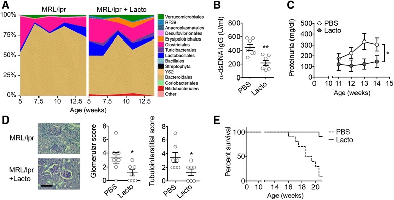
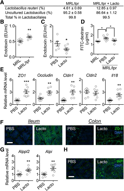
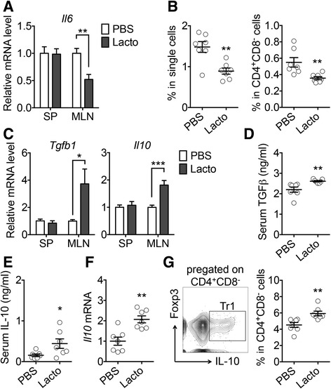
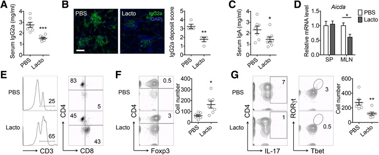
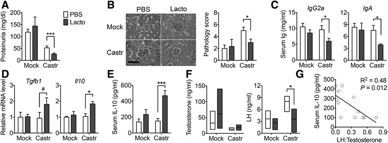
Results: Dysbiosis of gut microbiota promotes autoimmune disorders that damage extraintestinal organs. Here we report a role of gut microbiota in the pathogenesis of renal dysfunction in lupus. Using a classical model of lupus nephritis, MRL/lpr, we found a marked depletion of Lactobacillales in the gut microbiota. Increasing Lactobacillales in the gut improved renal function of these mice and prolonged their survival. We used a mixture of 5 Lactobacillus strains (Lactobacillus oris, Lactobacillus rhamnosus, Lactobacillus reuteri, Lactobacillus johnsonii, and Lactobacillus gasseri), but L. reuteri and an uncultured Lactobacillus sp. accounted for most of the observed effects. Further studies revealed that MRL/lpr mice possessed a "leaky" gut, which was reversed by increased Lactobacillus colonization. Lactobacillus treatment contributed to an anti-inflammatory environment by decreasing IL-6 and increasing IL-10 production in the gut. In the circulation, Lactobacillus treatment increased IL-10 and decreased IgG2a that is considered to be a major immune deposit in the kidney of MRL/lpr mice. Inside the kidney, Lactobacillus treatment also skewed the Treg-Th17 balance towards a Treg phenotype. These beneficial effects were present in female and castrated male mice, but not in intact males, suggesting that the gut microbiota controls lupus nephritis in a sex hormone-dependent manner.

Conclusions: This work demonstrates essential mechanisms on how changes of the gut microbiota regulate lupus-associated immune responses in mice. Future studies are warranted to determine if these results can be replicated in human subjects.

Keywords: Autoimmunity; Gut microbiota; Leaky gut; Lupus.


Conflict of interest statement

Ethics approval

This study was carried out in strict accordance with the recommendations in the Guide for the Care and Use of Laboratory Animals of the National Institutes of Health. The protocol was approved by the Institutional Animal Care and Use Committee (IACUC) of Virginia Tech College of Veterinary Medicine (Animal Welfare Assurance Number: A3208-01). For anesthesia and euthanasia, isoflurane and CO2 were used, respectively, according to the IACUC protocol.

Consent for publication

Not applicable.

Competing interests

The authors declare that they have no competing interests.

Publisher’s Note

Springer Nature remains neutral with regard to jurisdictional claims in published maps and institutional affiliations.

Figures

Fig. 1
Fig. 1
Lactobacillus spp. protect female lpr mice from LN. a Time-dependent changes of fecal microbiota upon PBS or Lactobacillus (Lacto) treatment (n = 4 mice per group). b Level of anti-dsDNA IgG in the blood of 10-week-old mice (n = 7 mice per group; **P < 0.01). c Level of proteinuria over time (n = 7 mice per group; paired t test; *P < 0.05). d Renal histopathology at 14 weeks of age (n = 7 mice per group; chi-square test; *P < 0.05). Left: PAS-stained kidney sections; bar equals 100 μm. e Survival rate (n = 10 mice per group; chi-square test; ****P < 0.0001)
Fig. 2
Fig. 2
Lactobacillus spp. restore gut mucosal barrier function in female lpr mice. a Percentage of Lactobacillus strains in the order Lactobacillales (n = 4 per group). b Level of endotoxin in the blood of 6-week-old lpr mice (n = 6 mice per group; **P < 0.01). c Level of endotoxin in the blood of 10-week-old lpr mice with or without Lactobacillus treatment (n = 6 or 7 mice per group; *P < 0.05). d Level of FITC-dextran diffused to the blood (n = 5 or 7 mice per group; *P < 0.05). e Transcript levels of tight junction proteins and IL-18 in intestinal epithelial cells of 14-week-old lpr mice (n = 7 mice per group; **P < 0.01, ***P < 0.005). f Immunohistochemical stains of ZO-1 (green) in the ileum or colon. Nuclear stain (DAPI) is shown in blue. Bar equals 75 μm. g Transcript levels of IAP genes in the epithelium (n = 7 mice per group; **P < 0.01). h Immunohistochemical stains of IAP (green) in the ileum. Bar equals 75 μm
Fig. 3
Fig. 3
Control of intestinal inflammation by gut microbiota in female lpr mice. a Transcript level of IL-6 in the spleen (SP) and MLN (n = 7 mice per group; **P < 0.01). b Percentage of IL-6-expressing cells in the MLN (n = 7 mice per group; **P < 0.01). c Transcript levels of TGFβ and IL-10 (n = 7 mice per group; *P < 0.05, ***P < 0.005). d Serum level of TGFβ (n = 7 mice per group; **P < 0.01). e Serum level of IL-10 (n = 7 mice per group; *P < 0.05). f Transcript level of IL-10 in the kidney (n = 7 mice per group; **P < 0.01). g FACS analysis of IL-10-expressing Tr1 cells in the MLN. Percentages of Tr1 cells in CD4+CD8 cells are shown (n = 7 mice per group; **P < 0.01)
Fig. 4
Fig. 4
Control of renal inflammation by gut microbiota in female lpr mice. a Serum level of IgG2a (n = 7 mice per group; ***P < 0.005). b Immunohistochemical stains of IgG2a (green) in the kidney. Bar equals 75 μm. Pathological scores are shown on the right (n = 4 mice per group; **P < 0.01). c Serum level of IgA (n = 7 or 8 mice per group; *P < 0.05). d Transcript level of Aicda (n = 7 mice per group; *P < 0.05). e Percentages of T cells and subpopulations in the kidney. f Percentage of CD4+Foxp3+ Treg cells in the kidney. Absolute Treg cell numbers are shown on the right (n = 7 mice per group; *P < 0.05). g FACS analysis of IL-17-producing CD4+ cells and percentage of RORγT+Tbet+ pathogenic Th17 cells in the kidney. Absolute pathogenic Th17 cell numbers are shown on the right (n = 7 mice per group; **P < 0.01)
Fig. 5
Fig. 5
Sex hormones and gut microbiota cooperatively regulate LN. a Proteinuria after surgery (Mock vs. Castr/castration) and treatment (PBS vs. Lacto) of male lpr mice (n = 5 mice per group; Mann-Whitney test; ***P < 0.005). b Renal histopathology (n = 5 mice per group; Mann-Whitney test; *P < 0.05). Left: PAS-stained kidney sections; bar equals 100 μm. c Serum levels of IgG2a and IgA (n = 5 mice per group; *P < 0.05). d Transcript levels of TGFβ and IL-10 in the MLN (n = 5 mice per group; #P < 0.1, *P < 0.05). e Serum level of IL-10 (n = 5 mice per group; ***P < 0.005). f Levels of testosterone and luteinizing hormone (LH) in the blood (n = 3 mice per group; *P < 0.05). g Negative correlation between serum IL-10 and the ratio of LH to testosterone

References

    1. Tsokos GC. Systemic lupus erythematosus. N Engl J Med. 2011;365(22):2110–21. doi: 10.1056/NEJMra1100359. - DOI - PubMed
    1. Lech M, Anders HJ. The pathogenesis of lupus nephritis. J Am Soc Nephrol. 2013;24(9):1357–66. doi: 10.1681/ASN.2013010026. - DOI - PMC - PubMed
    1. Tektonidou MG, Wang Z, Dasgupta A, Ward MM. Burden of serious infections in adults with systemic lupus erythematosus: a national population-based study, 1996–2011. Arthritis Care Res (Hoboken) 2015;67(8):1078–85. doi: 10.1002/acr.22575. - DOI - PMC - PubMed
    1. Mu Q, Zhang H, Luo XM. SLE: another autoimmune disorder influenced by microbes and diet? Front Immunol. 2015;6:608. doi: 10.3389/fimmu.2015.00608. - DOI - PMC - PubMed
    1. Hevia A, Milani C, Lopez P, Cuervo A, Arboleya S, Duranti S, et al. Intestinal dysbiosis associated with systemic lupus erythematosus. MBio. 2014;5(5):e01548–14. doi: 10.1128/mBio.01548-14. - DOI - PMC - PubMed

Publication types

MeSH terms