. 2017 Jul 11;11(1):15.
doi: 10.1186/s40246-017-0112-8.

Inferring clonal structure in HTLV-1-infected individuals: towards bridging the gap between analysis and visualization

Affiliations

Inferring clonal structure in HTLV-1-infected individuals: towards bridging the gap between analysis and visualization

Amir Farmanbar et al. Hum Genomics. .

Abstract

Background: Human T cell leukemia virus type 1 (HTLV-1) causes adult T cell leukemia (ATL) in a proportion of infected individuals after a long latency period. Development of ATL is a multistep clonal process that can be investigated by monitoring the clonal expansion of HTLV-1-infected cells by isolation of provirus integration sites. The clonal composition (size, number, and combinations of clones) during the latency period in a given infected individual has not been clearly elucidated.

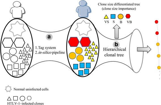
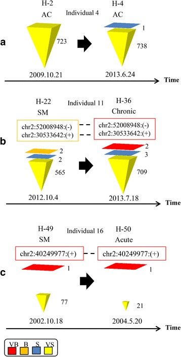
Methods: We used high-throughput sequencing technology coupled with a tag system for isolating integration sites and measuring clone sizes from 60 clinical samples. We assessed the role of clonality and clone size dynamics in ATL onset by modeling data from high-throughput monitoring of HTLV-1 integration sites using single- and multiple-time-point samples.

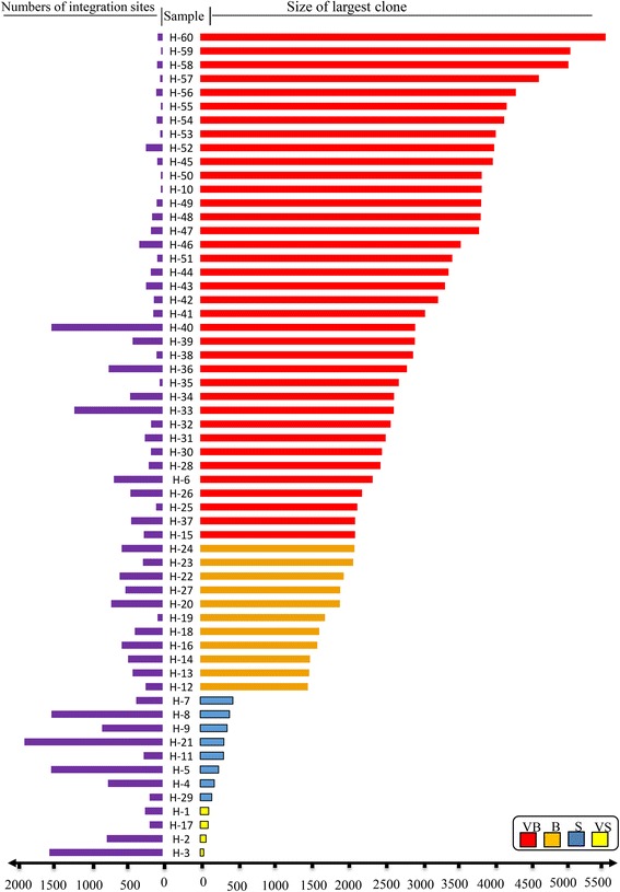
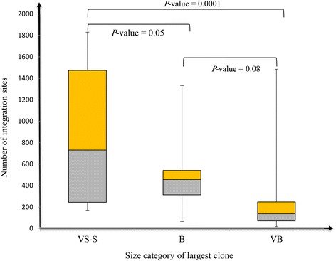
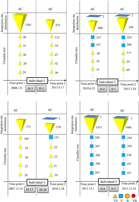
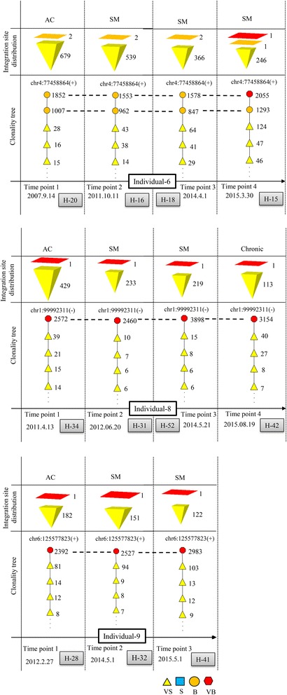
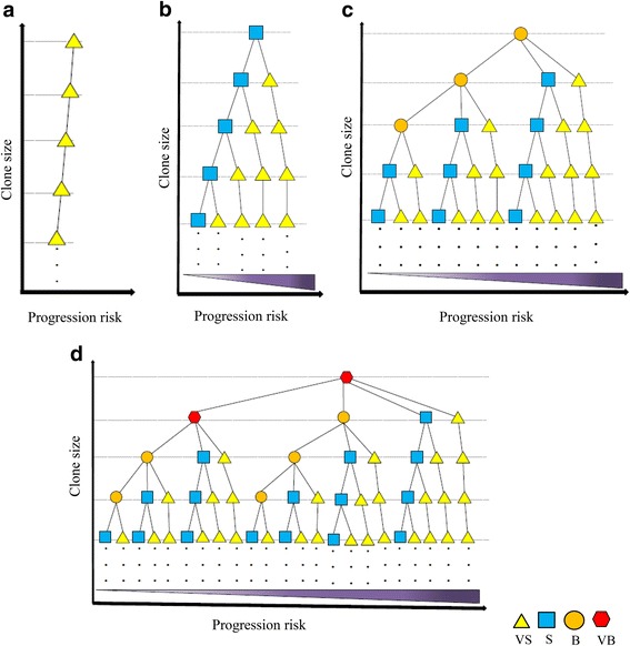
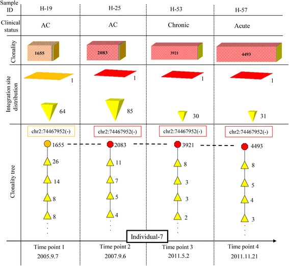
Results: From four size categories analyzed, we found that big clones (B; 513-2048 infected cells) and very big clones (VB; >2048 infected cells) had prognostic value. No sample harbored two or more VB clones or three or more B clones. We examined the role of clone size, clone combination, and the number of integration sites in the prognosis of infected individuals. We found a moderate reverse correlation between the total number of clones and the size of the largest clone. We devised a data-driven model that allows intuitive representation of clonal composition.

Conclusions: This integration site-based clonality tree model represents the complexity of clonality and provides a global view of clonality data that facilitates the analysis, interpretation, understanding, and visualization of the behavior of clones on inter- and intra-individual scales. It is fully data-driven, intuitively depicts the clonality patterns of HTLV-1-infected individuals and can assist in early risk assessment of ATL onset by reflecting the prognosis of infected individuals. This model should assist in assimilating information on clonal composition and understanding clonal expansion in HTLV-1-infected individuals.

Keywords: Adult T cell leukemia; Clonal expansion; Data-driven modeling; High-throughput sequencing; Human T cell leukemia virus type 1; Integration site; Prognostic indicator.


Conflict of interest statement

Competing interests

The authors declare that they have no competing interests.

Publisher’s Note

Springer Nature remains neutral with regard to jurisdictional claims in published maps and institutional affiliations.

Figures

Fig. 1
Fig. 1
Overview of clonality analysis and visualization of results. The workflow consists of two main bottlenecks. In bottleneck a, our original tag system was used to isolate HTLV-1 integration sites in infected individuals and measure the size of clones. Subsequently, our in silico pipeline was used to analyze the high-throughput sequencing data. In bottleneck b, which is the main focus of this manuscript, the observed clone sizes were classified and visualized using different shapes and colors using the concept of hierarchical tree structure
Fig. 2
Fig. 2
Clone size and integration sites in three representative individuals. Individuals are indicated at the top, followed by sample numbers and disease states at times 1 and 2. The number of integration sites is indicated beside color-coded representations of clone size. Clones with identical integration sites are shown in boxes color-coded for clone size and connected by dashed lines. a Individual 4 remained an asymptomatic carrier (AC) over time and had a large number of integration sites that fluctuated over time. b Individual 11 progressed from smoldering (SM) to chronic ATL. The integration sites of the two largest clones were identical over time. c Individual 16 progressed from SM to acute ATL. The integration site of the largest clone was identical over time. Results for other samples are shown in Additional file 2: Figure S3
Fig. 3
Fig. 3
Correlation between number of integration sites and the size of the largest clone across all samples. A moderate negative correlation was observed (R = –0.55 based on Pearson correlation coefficient)
Fig. 4
Fig. 4
Number of clones belonging to size categories across analyzed samples. Samples were divided into three groups (VS-S, B, and VB) based on the category of the largest clone. The number of integration sites isolated from each group was then plotted. P values were calculated by Student’s t test
Fig. 5
Fig. 5
Clonality data of ACs who remained ACs. High numbers of integration sites were detected from each sample. Only S and VS clones were observed
Fig. 6
Fig. 6
Clonality data of ACs who progressed to ATL. Clones with identical integration sites are connected by horizontal dashed lines. B or VB clones were detected in addition to VS and/or S clones, and their integration sites were constant over time
Fig. 7
Fig. 7
ad Global scheme of clonality dynamics among analyzed samples represented by tree structures. Each tree begins with a root of a different clone size (VS, S, B, and VB). The clonality status of each sample can be presented by an individual path. Within each tree, clone size decreases from the root to the leaves. In addition, samples on the right side of a tree have a greater risk of disease progression compared with those on the left. Thus, clone size, order, and disease progression of each sample can be compared to the other samples
Fig. 8
Fig. 8
Clonality tree for a patient (individual 7) monitored over 6 years who progressed from AC status to ATL onset. The patient had a B clone at time point 1 and a VB clone at time points 2, 3, and 4. The integration site of the largest clone was identical across all time points, as indicated by the dashed horizontal lines. Clonality of this high-risk sample was notably different from that of a typical AC

Similar articles

Cited by

References

    1. Nowell PC. The clonal evolution of tumor cell populations. Science. 1976;194:23–28. doi: 10.1126/science.959840. - DOI - PubMed
    1. Crespi B, Summers K. Evolutionary biology of cancer. Trends Ecol Evol. 2005;20:545–552. doi: 10.1016/j.tree.2005.07.007. - DOI - PubMed
    1. Merlo LMF, Pepper JW, Reid BJ, Maley CC. Cancer as an evolutionary and ecological process. Nat Rev Cancer. 2006;6:924–935. doi: 10.1038/nrc2013. - DOI - PubMed
    1. Greaves M. Evolutionary determinants of cancer. Cancer Discov. 2015;5:806–820. doi: 10.1158/2159-8290.CD-15-0439. - DOI - PMC - PubMed
    1. Sun YJ, Yao J, Nowak NJ, Goodison S. Cancer progression modeling using static sample data. Genome Biol. 2014;15:440. DOI:10.1186/s13059-014-0440-0. - PMC - PubMed

Publication types

LinkOut - more resources