. 2017 Aug 4;429(16):2449-2462.
doi: 10.1016/j.jmb.2017.07.003. Epub 2017 Jul 10.

Molecular Origins of the Compatibility between Glycosaminoglycans and Aβ40 Amyloid Fibrils

Affiliations

Molecular Origins of the Compatibility between Glycosaminoglycans and Aβ40 Amyloid Fibrils

Katie L Stewart et al. J Mol Biol. .

Abstract

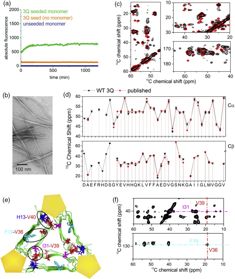
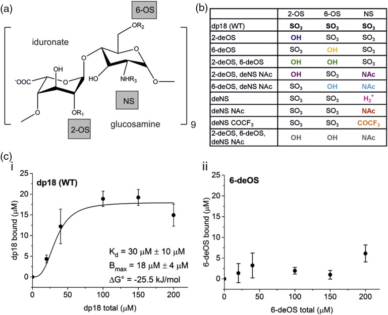
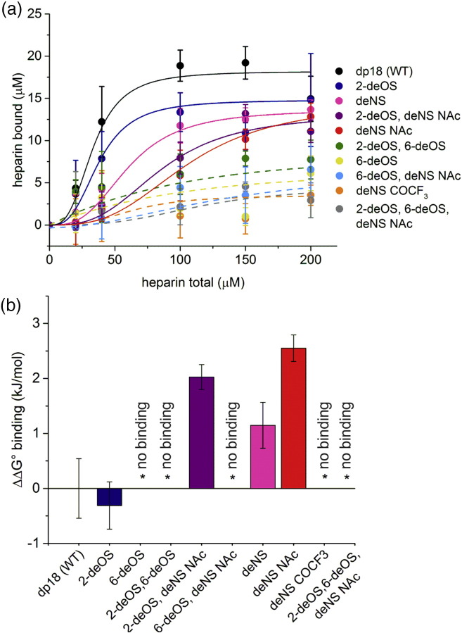
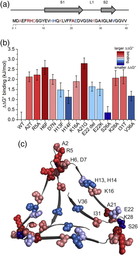
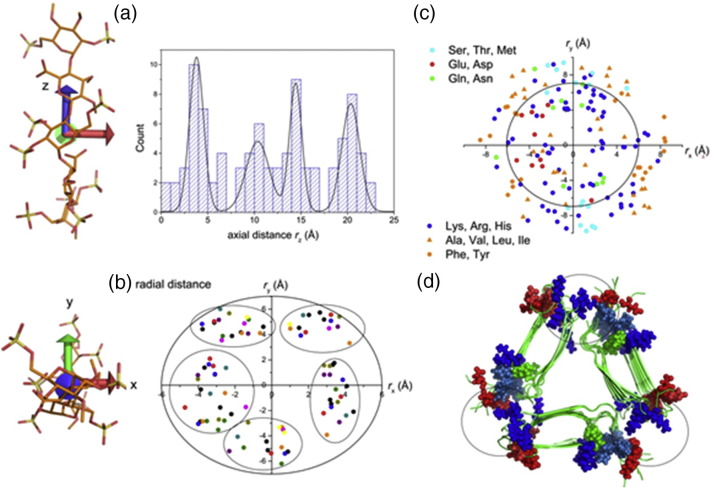
The Aβ peptide forms extracellular plaques associated with Alzheimer's disease. In addition to protein fibrils, amyloid plaques also contain non-proteinaceous components, including glycosaminoglycans (GAGs). We have shown previously that the GAG low-molecular-weight heparin (LMWH) binds to Aβ40 fibrils with a three-fold-symmetric (3Q) morphology with higher affinity than Aβ40 fibrils in alternative structures, Aβ42 fibrils, or amyloid fibrils formed from other sequences. Solid-state NMR analysis of the GAG-3Q fibril complex revealed an interaction site at the corners of the 3Q fibril structure, but the origin of the binding specificity remained obscure. Here, using a library of short heparin polysaccharides modified at specific sites, we show that the N-sulfate or 6-O-sulfate of glucosamine, but not the 2-O-sulfate of iduronate within heparin is required for 3Q binding, indicating selectivity in the interactions of the GAG with the fibril that extends beyond general electrostatic complementarity. By creating 3Q fibrils containing point substitutions in the amino acid sequence, we also show that charged residues at the fibril three-fold apices provide the majority of the binding free energy, while charged residues elsewhere are less critical for binding. The results indicate, therefore, that LMWH binding to 3Q fibrils requires a precise molecular complementarity of the sulfate moieties on the GAG and charged residues displayed on the fibril surface. Differences in GAG binding to fibrils with distinct sequence and/or structure may thus contribute to the diverse etiology and progression of amyloid diseases.

Keywords: Alzheimer's disease; amyloid fibrils; amyloid β; glycosaminoglycans; heparin binding.


Figures

Image 1
Graphical abstract
Fig. 1
Fig. 1
Aβ40 fibrils of 3Q morphology are produced by seeded growth. (a) Seeded growth 5% (v/v) of 3Q fibril seeds with Aβ40 WT monomers (20 μM) monitored by ThT fluorescence. (b) TEM image of fibrils formed in panel A after 24 h. The scale bar represents 200 nm. (c) Three regions of a 2D 13C–13C SSNMR spectrum (with 50-ms DARR mixing) of Aβ40 fibrils prepared with 3Q seeding. The experimental spectrum (black) is overlaid with a simulated spectrum (red) based on the reported 13C chemical shifts for the 3Q morphology . (d) 13C Cα and Cβ chemical shifts measured from the spectrum in panel C compared with previous published assignments by Paravastu and colleagues . Error bars on the current data represent ± half the line widths measured at half peak height, with an average of 1.07 ppm across the entire sequence. (e) Structure of the 3Q model of Aβ40 (viewed down the fibril axis) based on SSNMR restraints (PDB ID: 2LMQ[45]), showing the previously determined LMWH binding sites (yellow pentagons). Circled regions highlight three long-range couplings between residues observed in Ref. , which are diagnostic of the hairpin structure (H13–V40, F19–V36) and of the quaternary packing arrangement in the 3Q morphology (I31–V39). (f) Regions of a 2D 13C–13C SSNMR spectrum (200-ms DARR mixing) showing long-range F19–V36 and I31–V39 cross peaks. Spectra are shown in Fig. S2 and assignments in Table S1.
Fig. 2
Fig. 2
Modifications of heparin substituents affect binding to the 3Q fibril. (a) Structure of a heparin disaccharide unit of dp18 with labeled sulfate groups. (b) List of modifications to the heparin structure based on panel A, with modifications shown in color. (c) Binding curves of modified heparins to 3Q fibrils of Aβ40. Each data point represents the average of three replicates with standard deviation at each heparin concentration. Bmax, Kd, and ΔG° binding values are reported where binding was detected. The solid line, where applicable, was obtained by non-linear least-squares fitting of a Hill function. Additional heparin variant binding data are shown in Fig. S3 and Table S2.
Fig. 3
Fig. 3
Summary of heparin variants binding to 3Q fibrils of Aβ40. (a) Binding of 3Q fibrils to all modified heparin constructs tested overlaid for comparison. Solid lines indicate heparin variants for which binding could be determined. Dashed lines indicate heparin variants that show little to no binding to 3Q fibrils and for which a Kd value could not be determined. Lines were obtained by non-linear least-squares fitting of a Hill function. Colors correspond to Fig. 2b. (b) Comparison of ΔΔG° binding of modified heparins to 3Q fibrils, relative to unmodified heparin (dp18), shown in Fig. 2c-i. Asterisks denote heparin variants that showed little or no binding. Error bars depict the standard deviation over three replicate assays.
Fig. 4
Fig. 4
Binding of 3Q fibril variants H6F, K16A, H13F, and S26A to LMWH shows a range of binding free energies. (a) ThT fluorescence confirms that 3Q seeds from WT Aβ40 can be used to seed fibril formation of Aβ40 variants. Seed [5% (v/v)] was added to each monomeric variant at or near the start of incubation, as indicated by an arrow on each panel, causing a rapid increase in fluorescence (red or blue). Each curve shown is a representative based on four replicates. (b) TEM confirms the presence of fibrils after 24 h of seeded growth. (c) Binding curves of variant 3Q fibrils to LMWH indicate a range of binding free energies. Each data point represents the average of three replicates (with standard deviation) at each LMWH concentration. The solid line was obtained by non-linear least-squares fitting of a Hill function. Additional binding data are found in Fig. S7 and Table S4. Colors of the panels correspond to the ΔΔG° binding gradient used in Fig. 5.
Fig. 5
Fig. 5
Single-substitution variants highlight the importance of the 3Q fibril “corners” in LMWH binding. (a) The sequence of Aβ40 with the locations of strands and substitution positions in the 3Q morphology shown. (Note that recombinant Aβ40 contains an N-terminal methionine residue at position 0.) (b) Comparison of ΔΔG° values obtained from the binding assay for 3Q variants, relative to WT 3Q fibrils, colored from smaller (blue) to larger (red) changes in ΔΔG° binding. Error bars depict variation in binding between three replicate assays. (c) The Aβ40 3Q fibril structure (from PDBID 2LMQ with an added disordered N-terminus) with substituted residues mapped as space-filling models, colored as in parts A and B.
Fig. 6
Fig. 6
Rules for heparin–fibril binding. Crystal structures of 16 unrelated proteins (Table S5) containing bound heparin fragments ranging from dimers to hexamers were compiled and any extraneous molecules were removed, leaving the protein and heparin. The structures were then rotated into a new reference frame corresponding to the principal axes of inertia of heparin in each case, with the origin at the center of mass of heparin. (a) The distribution of sulfate groups along the z-axis. (b) Cross-section view of the heparin molecules in which each point represents the position of a sulfur atom. (c) The positions of amino acid residues within 4 Å of one or more sulfate groups. (d) The structure of 3Q fibrils (2LMQ [45]) with acidic and basic residues shown in red and blue, respectively. The circles represent the cross-sectional space of heparin.

Similar articles

Cited by

References

    1. Sipe J.D., Benson M.D., Buxbaum J.N., Ikeda S., Merlini G., Saraiva M.J.M. Amyloid fibril proteins and amyloidosis: chemical identification and clinical classification, International Society of Amyloidosis 2016 nomenclature guidelines. Amyloid. 2016;23:209–213. - PubMed
    1. Eichner T., Radford S.E. A diversity of assembly mechanisms of a generic amyloid fold. Mol. Cell. 2011;43:8–18. - PubMed
    1. Chiti F., Dobson C.M. Protein misfolding, amyloid formation, and human disease: a summary of progress over the last decade. Annu. Rev. Biochem. 2017 - PubMed
    1. Bradley-Whitman M.A., Timmons M.D., Beckett T.L., Murphy M.P., Lynn B.C., Lovell M.A. Nucleic acid oxidation: an early feature of Alzheimer's disease. J. Neurochem. 2014;128:294–304. - PMC - PubMed
    1. Han S., Kollmer M., Markx D., Claus S., Walther P., Fandrich M. Amyloid plaque structure and cell surface interactions of beta-amyloid fibrils revealed by electron tomography. Sci. Rep. 2017;7:43577. - PMC - PubMed

Publication types

MeSH terms