

Longitudinal characterisation of function and structure of Bietti crystalline dystrophy: report on a novel homozygous mutation in CYP4V2
- PMID: 28698241
- PMCID: PMC6208130
- DOI: 10.1136/bjophthalmol-2016-309696
Longitudinal characterisation of function and structure of Bietti crystalline dystrophy: report on a novel homozygous mutation in CYP4V2
Abstract
Background: Bietti crystalline dystrophy (BCD) is a rare inherited disorder characterised by fine crystalline deposits in the corneal limbus and retinal posterior pole. In 2004, mutations in the CYP4V2 gene were identified as the cause of BCD. Here, we describe the report of a homozygous point mutation in a patient with BCD and provide detailed characterisation of functional and structural changes over 20 years.
Methods: At regular intervals, the patient underwent repeat ophthalmic evaluations. DNA was extracted from buccal swabs, amplified by standard PCR and analysed for homology to the CYP4V2 sequence. Homology modelling was conducted using Iterative Threading ASSEmbly Refinement and molecular dynamics simulations using GROningen MAchine for Chemical Simulations.
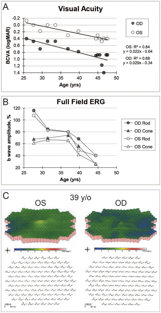
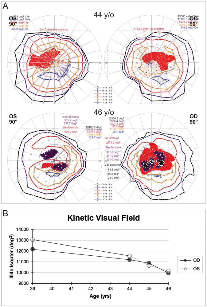
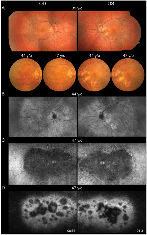
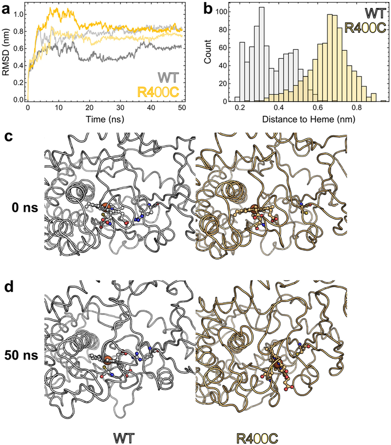
Results: The proband, a 47-year-old woman of German ancestry was diagnosed with crystalline retinopathy at age 25. Over the next 20 years, visual acuity and function gradually declined with progression of retinal pigment epithelium and choroidal atrophy. When first tested at 39 years of age, the multifocal electroretinogram (ERG) was markedly abnormal, more so for the right eye, whereas the full-field ERG was more symmetrical and lagged other measures of visual function. Gene sequencing showed a single C>T point mutation in exon 9 encoding a R400C amino acid change. Computational modelling suggests the mutation impairs function due to loss of a hydrogen bonding interaction with the propionate side chains of the haeme prosthetic group.
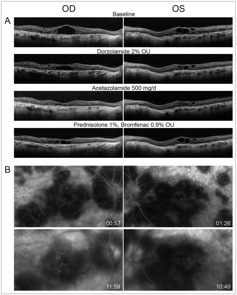
Conclusion: This is the first report of a homozygous R400C mutation in CYP4V2 with protein modelling showing high likelihood of enzyme dysfunction. The comprehensive long-term clinical follow-up provides insight into disease progression and highlights possible anti-inflammatory modulation of disease severity.
Keywords: Bietti crystalline dystrophy; CYP4V2; macular edema; protein modeling.
© Article author(s) (or their employer(s) unless otherwise stated in the text of the article) 2018. All rights reserved. No commercial use is permitted unless otherwise expressly granted.
Conflict of interest statement
Competing interests: Weleber is a consultant to Novartis, Pfizer and Wellstat; is a member of the scientific advisory board for Applied Genetic Technologies Corp; and serves on the scientific advisory board for the Foundation Fighting Blindness (the relationship has been reviewed and managed by Oregon Health and Science University). Dr Weleber also reports having received grants and personal fees from the Foundation Fighting Blindness and Applied Genetic Technologies Corp and other support from Sanofi, all outside the submitted work. In addition, Dr Weleber has a patent (US patent 8 657 446, method and apparatus for visual field monitoring, also known as Visual Field Monitoring and Analysis, or VFMA, which has not been issued).
Figures
Similar articles
-
Detailed functional and structural phenotype of Bietti crystalline dystrophy associated with mutations in CYP4V2 complicated by choroidal neovascularization.Ophthalmic Genet. 2016 Dec;37(4):445-452. doi: 10.3109/13816810.2015.1126616. Epub 2016 Mar 30. Ophthalmic Genet. 2016. PMID: 27028354 Free PMC article.
-
Genotype and Long-term Clinical Course of Bietti Crystalline Dystrophy in Korean and Japanese Patients.Ophthalmol Retina. 2021 Dec;5(12):1269-1279. doi: 10.1016/j.oret.2021.02.009. Epub 2021 Feb 23. Ophthalmol Retina. 2021. PMID: 33636399
-
Identification of novel CYP4V2 genotypes associated with Bietti crystalline dystrophy and atypical anterior segment phenotypes in Spanish patients.Acta Ophthalmol. 2018 Nov;96(7):e865-e873. doi: 10.1111/aos.13768. Epub 2018 Apr 25. Acta Ophthalmol. 2018. PMID: 29691984
-
Genetics of Bietti Crystalline Dystrophy.Asia Pac J Ophthalmol (Phila). 2016 Jul-Aug;5(4):245-52. doi: 10.1097/APO.0000000000000209. Asia Pac J Ophthalmol (Phila). 2016. PMID: 27228076 Review.
-
Unravelling CYP4V2: Clinical features, genetic insights, pathogenic mechanisms and therapeutic strategies in Bietti crystalline corneoretinal dystrophy.Prog Retin Eye Res. 2025 Jul;107:101377. doi: 10.1016/j.preteyeres.2025.101377. Epub 2025 Jun 5. Prog Retin Eye Res. 2025. PMID: 40482806 Review.
Cited by
-
Evaluating precision medicine approaches for gene therapy in patient-specific cellular models of Bietti crystalline dystrophy.JCI Insight. 2024 Aug 22;9(16):e177231. doi: 10.1172/jci.insight.177231. JCI Insight. 2024. PMID: 39171529 Free PMC article.
-
Natural history of progressive vision loss in Bietti crystalline dystrophy: a model-based meta-analysis.BMJ Open Ophthalmol. 2025 Apr 12;10(1):e001908. doi: 10.1136/bmjophth-2024-001908. BMJ Open Ophthalmol. 2025. PMID: 40221146 Free PMC article.
-
Expanding the Phenotypic and Genotypic Spectrum of Bietti Crystalline Dystrophy.Genes (Basel). 2021 May 10;12(5):713. doi: 10.3390/genes12050713. Genes (Basel). 2021. PMID: 34068831 Free PMC article.
-
Application of Electrophysiology in Non-Macular Inherited Retinal Dystrophies.J Clin Med. 2023 Nov 6;12(21):6953. doi: 10.3390/jcm12216953. J Clin Med. 2023. PMID: 37959417 Free PMC article. Review.
-
CYP4V2 mutation screening in an Iranian Bietti crystalline dystrophy pedigree and evidence for clustering of CYP4V2 mutations.J Curr Ophthalmol. 2019 Mar 2;31(2):172-179. doi: 10.1016/j.joco.2019.01.007. eCollection 2019 Jun. J Curr Ophthalmol. 2019. PMID: 31317096 Free PMC article.
References
-
- Ng DS, Lai TY, Ng TK, et al. Genetics of Bietti crystalline dystrophy. Asia Pac J Ophthalmol 2016;5:245–52. - PubMed
Publication types
MeSH terms
Substances
Supplementary concepts
Grants and funding
LinkOut - more resources
Full Text Sources
Other Literature Sources
Medical