. 2017 Jul 25;114(30):8029-8034.
doi: 10.1073/pnas.1703406114. Epub 2017 Jul 11.

Deletion of Gas2l3 in mice leads to specific defects in cardiomyocyte cytokinesis during development

Affiliations

Deletion of Gas2l3 in mice leads to specific defects in cardiomyocyte cytokinesis during development

Sabine Stopp et al. Proc Natl Acad Sci U S A. .

Abstract

GAS2L3 is a recently identified cytoskeleton-associated protein that interacts with actin filaments and tubulin. The in vivo function of GAS2L3 in mammals remains unknown. Here, we show that mice deficient in GAS2L3 die shortly after birth because of heart failure. Mammalian cardiomyocytes lose the ability to proliferate shortly after birth, and further increase in cardiac mass is achieved by hypertrophy. The proliferation arrest of cardiomyocytes is accompanied by binucleation through incomplete cytokinesis. We observed that GAS2L3 deficiency leads to inhibition of cardiomyocyte proliferation and to cardiomyocyte hypertrophy during embryonic development. Cardiomyocyte-specific deletion of GAS2L3 confirmed that the phenotype results from the loss of GAS2L3 in cardiomyocytes. Cardiomyocytes from Gas2l3-deficient mice exhibit increased expression of a p53-transcriptional program including the cell cycle inhibitor p21. Furthermore, loss of GAS2L3 results in premature binucleation of cardiomyocytes accompanied by unresolved midbody structures. Together these results suggest that GAS2L3 plays a specific role in cardiomyocyte cytokinesis and proliferation during heart development.

Keywords: GAS2L3; binucleation; cardiomyocytes; cytokinesis.


Conflict of interest statement

The authors declare no conflict of interest.

Figures

Fig. S1.
Fig. S1.
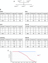
A conditional allele of Gas2l3. (A) Scheme of the conditional Gas2l3 allele. After removal of the selection cassette with flp recombinase, Exon 6 is flanked by loxP sites. Exon 6 can be removed by the activity of Cre recombinase. (B) Scheme of the position of primers used for genotyping of the different Gas2l3 alleles. The table shows the size of the PCR products for the different primer pairs. (C) Results of PCR genotyping with the indicated primer pairs. (D) Gas2l3 conditional MEFs expressing a CreER transgene were treated with 4-OH-tamoxifen to activate CreER. This Cre-activation resulted in deletion of Exon 6 as determined by PCR with primers specific for the fl and - allele. (E) Loss of Gas2l3 mRNA expression after Cre-mediated deletion as determined by RT-qPCR. (F) MEFs were treated with 4-OHT or with solvent (ethanol), and lysates were immunoprecipitated and immunoblotted with anti-GAS2L3 antibodies. HA-tagged GAS2L3 expressed in 293 cells served as a control. ****P < 0.0001.
Fig. S2.
Fig. S2.
Lethality of Gas2l3−/− mice. (A) Breeding scheme and resulting genotypes. (B) Result of the genotyping at the indicated developmental time points. Shown is the actual number of mice compared with the expected number of animals at normal Mendelian frequency. (C) Graph of viability of wild-type and Gas2l3−/− embryos and mice versus developmental stage.
Fig. 1.
Fig. 1.
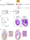
Postnatal lethality and dilated hearts of Gas2l3 knockout mice. (A) Fixed hearts from 43-d and 128-d-old animals. Wild-type (+/+) and heterozygote (+/−) mice served as controls. (B) Heart-to-body weight of 5- to 7-d-old and 14-d-old Gas2l3−/− and control mice. n > 10 mice per group. (C) H&E-stained sagittal sections demonstrating dilated ventricles in Gas2l3−/− animals compared with control mice. (D) H&E stained transverse sections of Gas2l3−/− and Gas2l3+/+ hearts. (E and F) Cardiac function was analyzed by echocardiography in 6-wk-old mice. (E) Representative images of echocardiographic analysis. (F) Quantification of LVAW and LVPW (left ventricular anterior and posterior wall thickness), LVEDD and LVESD left (ventricular end-diastolic and end-systolic dimensions) and fractional shortening (FS). n = 3–5 mice per group. (Scale bars: C, 0.5 mm; D, 1 mm; E, vertical 2 mm, horizontal 0.1 s.) *P < 0.05; **P < 0.01; ***P < 0.001; ****P < 0.0001; ns, not significant.
Fig. S3.
Fig. S3.
Histopathology of Gas2l3−/− mice at P10 compared with wild-type littermates. Hematoxylin-eosin (H&E)-stained sections of various organs from control mice and Gas2l3−/− mice revealed no difference in the organ histology between the control animals and Gas2l3 knockout animals. (Scale bars: 0.1 cm.)
Fig. 2.
Fig. 2.
Hypertrophy and fibrosis of Gas2l3 knockout hearts. (A) Immunofluorescence staining of cTNT in a wild-type and Gas2l3−/− heart at P5 (green). Nuclei were stained with Hoechst (blue). (B) WGA staining at P7 and P14. A quantification of the cross-sectional area is shown on the right. (C) Sirius red staining demonstrating fibrosis in Gas2l3−/− hearts. (D) mRNA expression of the indicated genes was analyzed by RT-qPCR in P7 and P14 hearts. Expression was normalized to 18S rRNA. n = 3 mice per group. (Scale bars: A, Left and Center, 75 µm; A, Right, 10 µm; B, 50 µm; C, 500 µm.) *P < 0.05; **P < 0.01; ***P < 0.001; ****P < 0.0001; ns, not significant.
Fig. S4.
Fig. S4.
The phenotype of Gas2l3 loss is cardiomyocyte-specific. (A) Scheme of cardiomyocyte-specific deletion of Gas2l3 by Nkx2.5-Cre. (B) Increased heart-to-body weight at P14 in cardiac-specific Gas2l3 knockout mice generated by crossing Nkx2.5-Cre mice with Gas2l3fl/fl mice. Data shown as mean ± SEM. Student’s t test. *P < 0.05. (C) H&E-stained section showing dilated cardiomyopathy of a 15-d-old Nkx2.5-Cre;Gas2l3fl/fl mouse compared with an age-matched control mouse. (Scale bar: 500 µm.) (D) Effect of cardiac specific deletion of Gas2l3 was assessed by sirius red staining.
Fig. S5.
Fig. S5.
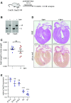
GAS2L3 is dispensable in the adult heart. (A) Scheme of inducible deletion of Gas2l3 in Gas2l3fl/fl; CreERT2 mice by injection of tamoxifen on days 12–14 (P12–P14) and analysis 14 wk later. (B) Deletion of Gas2l3 upon tamoxifen (tam) injection was confirmed by PCR of genomic DNA isolated from tail biopsies and from the heart. (C) Heart-to-body weight of mice treated without (-) or with (+) tamoxifen, 14 wk after tamoxifen injection. n = 7–9 mice per group. Mean with SEM. Student’s t test. (D) H&E and Sirius Red stained heart section 14 wk after deletion of Gas2l3 in Gas2l3fl/fl;CreERT2 mice compared with a heart from a littermate control animal. (E) mRNA expression of Gas2l3 was analyzed by RT-qPCR during heart development in E14.5 to P7 hearts. Expression was normalized to 18S rRNA. Gas2l3 mRNA is down-regulated at P2 when cardiomyocytes binucleate and exit from the cell cycle. n = 3 mice per group. One-way ANOVA with Bonferroni post hoc test. ****P < 0.0001; ns, not significant.
Fig. 3.
Fig. 3.
Decreased proliferation of Gas2l3 knockout cardiomyocytes. (A) Total number of cardiomyocytes in hearts from control mice and Gas2l3−/− mice at the indicated developmental time points. Cardiomyocytes were isolated, fixed, and quantified with a hematocytometer. (B) Area of fixed cardiomyocytes at the indicated time points. (C, Left) Representative immunofluorescence staining of Ki67 and cTnT in a E14.5 heart section. (Scale bars: 25 µm.) (Right) Quantification of Ki67-positive cardiomyocytes in E14.5, E16.5, E18.5, P1, and P7 hearts. ***P < 0.001; ****P < 0.0001; ns, not significant.
Fig. S6.
Fig. S6.
Proliferation and binucleation in the embryonic liver is unchanged in Gas2l3 knockout mice. (A) Representative immunofluorescence staining of Ki67 in E14.5 Gas2l3−/− and wild-type livers (Left). (Scale bars: 50 µm.) Quantification of Ki67-positive hepatocytes in E14.5 livers (Right). Student’s t test. (B) Quantification of binucleated hepatocytes in control and Gas2l3−/− mice at P10 and P128. Fisher’s exact test. ns, not significant.
Fig. 4.
Fig. 4.
RNA-Seq analysis of Gas2l3−/− hearts. (A) RNA-Seq was performed on RNA isolated from E18.5 wild-type and Gas2l3−/− hearts (n = 3 hearts per group). GSEA plot for the GO term mitosis. (B) GSEA identified a significant enrichment of targets of the p53 transcription factor in Gas2l3 knockout hearts. (C) Heatmaps of the p53 target genes enriched in Gas2l3−/− hearts compared with wild-type hearts. (D) RT-qPCR was used to confirm up-regulation of the indicated p53-target genes. (E) The levels of p21, p53, and hypophosphorylated pRB in lysates from wild-type (+/+) and Gas2l3−/− hearts at E18.5 were determined by immunoblotting. Tubulin served as a loading control. *P < 0.05;**P < 0.01; ***P < 0.001; ****P < 0.0001.
Fig. S7.
Fig. S7.
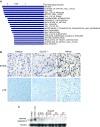
Genes with decreased expression in Gas2l3−/− hearts are linked to processes related to cell cycle regulation, mitosis, and chromosome segregation but loss of GAS2L3 does not result in apoptosis. (A) GSEA was used to identify the top 20 Gene Ontology (GO) terms associated with genes down-regulated in Gas2l3−/− hearts as identified by RNA-Seq (Fig. 4). (B) TUNEL staining of wild-type and Gas2l3−/− hearts at the indicated developmental time points indicating the absence of apoptosis. DNase I treatment served as a positive control. (C) Immunoblot analysis of cleaved caspase 3 as a marker of apoptotic cells. Doxorubicin (dox) treatment of H460 cells served as a positive control for the induction of apoptosis.
Fig. 5.
Fig. 5.
Defective cytokinesis in Gas2l3-deficient cardiomyocytes. (A and B) Quantification of mononucleated and binucleated cardiomoyocytes in control and Gas2l3−/− mice. (Inset) Example immunofluorescence images of mononucleated and binucleated cardiomyocytes stained with Connexin 43 and Hoechst are shown. (C) Representative immunofluorescence staining of phospho-histone H3 (Ser10) (pH3) and cTnT in an E14.5 heart section (Left). Quantification of pH3-positive cardiomyocytes in E14.5, E16.5, and E18.5 heart sections. (D) Representative confocal immunofluorescence image of Aurora B and cTnT in an E16.5 heart section (Left). Quantification of Aurora B-positive cardiomyocytes per section (Right). (E) E14.5 Gas2l3fl/fl;CreERT2 were treated for 96 h with 4-OHT and stained for WGA (green), Nkx2.5 (red), and Hoechst. Arrows point to binucleated cells. (F) Detection of unresolved midbody structures in Gas2l3fl/fl;CreERT2 cardiomyocytes by staining for Aurora B (red). Cardiomyocytes were identified by staining for cTnT (green). Nuclei were stained with Hoechst. (G) Quantification of binuclear E14.5 Gas2l3fl/fl;CreERT2 cardiomyocytes and cardiomyocytes with unresolved midbodies after treatment with or without 4-OHT for the indicated times. (H) Localization of GAS2L3 to the spindle midzone and the midbody of cardiomyocytes as detected by staining with an antiserum directed at murine GAS2L3 (red). Cardiomyocytes were identified by staining for Nkx2.5 (green). (Scale bars: A and B, Insets, C, Left, E, and F, 25 µm; D, Left, 75 µm; H, 5 µm.) **P < 0.01; ****P < 0.0001; ns, not significant.
Fig. S8.
Fig. S8.
Induction of Cre-recombinase in control cardiomyocytes does not lead to binucleation. (A) Cardiomyocytes isolated from E14.5 Gas2l3fl/fl;CreERT2 embryos were treated for 48 h with and without 4-OHT. Deletion of Gas2l3 was confirmed by genomic PCR. (B) Cardiomyocytes isolated from E14.5 Gas2l3+/+;CreERT2 embryos were treated for 48–72 h with and without 4-OHT and stained for WGA (green), Nkx2.5 (red), and Hoechst. (Scale bars: 25 µm.) (C) Quantification of binuclear E14.5 Gas2l3+/+;CreERT2 cardiomyocytes treated with 4-OHT for the indicated time points. P values were determined by χ2 test.
Fig. S9.
Fig. S9.
Localization of Aurora B, Anillin, and Myosin II to the midbody upon deletion of Gas2l3. (A) Localization of Aurora B (red) and Anillin (magenta) in untreated and 4-OHT–treated Gas2l3fl/fl;CreERT2 cardiomyocytes. Cardiomyocytes were identified by staining for cTnT (green). Nuclei were counterstained with Hoechst. (B) Localization of Myosin IIB (red) and Aurora B (magenta) in untreated and 4-OHT–treated Gas2l3fl/fl;CreERT2 cardiomyocytes. Cardiomyocytes were identified by staining for cTnT (green). Nuclei were stained with Hoechst. (Scale bars: 25 µm.)

Comment in

  • GAS2L3: Coordinator of cardiomyocyte cytokinesis?
    Gründl M, Engel FB, Gaubatz S. Gründl M, et al. Cell Cycle. 2017 Oct 18;16(20):1853-1854. doi: 10.1080/15384101.2017.1372546. Epub 2017 Sep 22. Cell Cycle. 2017. PMID: 28937871 Free PMC article. No abstract available.

References

    1. Li F, Wang X, Capasso JM, Gerdes AM. Rapid transition of cardiac myocytes from hyperplasia to hypertrophy during postnatal development. J Mol Cell Cardiol. 1996;28:1737–1746. - PubMed
    1. Soonpaa MH, Kim KK, Pajak L, Franklin M, Field LJ. Cardiomyocyte DNA synthesis and binucleation during murine development. Am J Physiol. 1996;271:H2183–H2189. - PubMed
    1. van Amerongen MJ, Engel FB. Features of cardiomyocyte proliferation and its potential for cardiac regeneration. J Cell Mol Med. 2008;12:2233–2244. - PMC - PubMed
    1. Ahuja P, Sdek P, MacLellan WR. Cardiac myocyte cell cycle control in development, disease, and regeneration. Physiol Rev. 2007;87:521–544. - PMC - PubMed
    1. Zebrowski DC, Engel FB. The cardiomyocyte cell cycle in hypertrophy, tissue homeostasis, and regeneration. Rev Physiol Biochem Pharmacol. 2013;165:67–96. - PubMed

Publication types

Substances