. 2017 Aug 16;37(33):7824-7836.
doi: 10.1523/JNEUROSCI.0691-17.2017. Epub 2017 Jul 11.

Delayed Cryptochrome Degradation Asymmetrically Alters the Daily Rhythm in Suprachiasmatic Clock Neuron Excitability

Affiliations

Delayed Cryptochrome Degradation Asymmetrically Alters the Daily Rhythm in Suprachiasmatic Clock Neuron Excitability

Sven Wegner et al. J Neurosci. .

Abstract

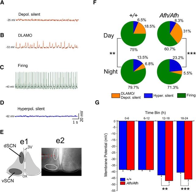
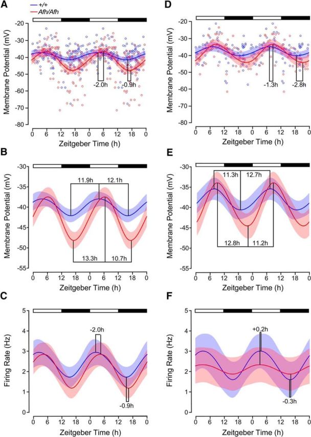
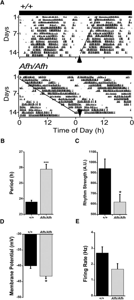
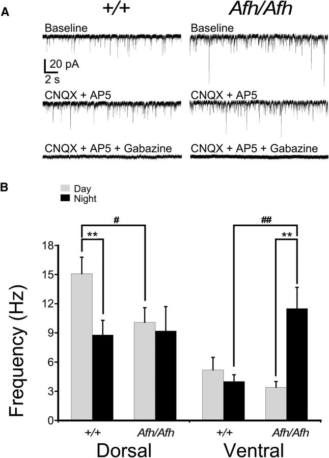
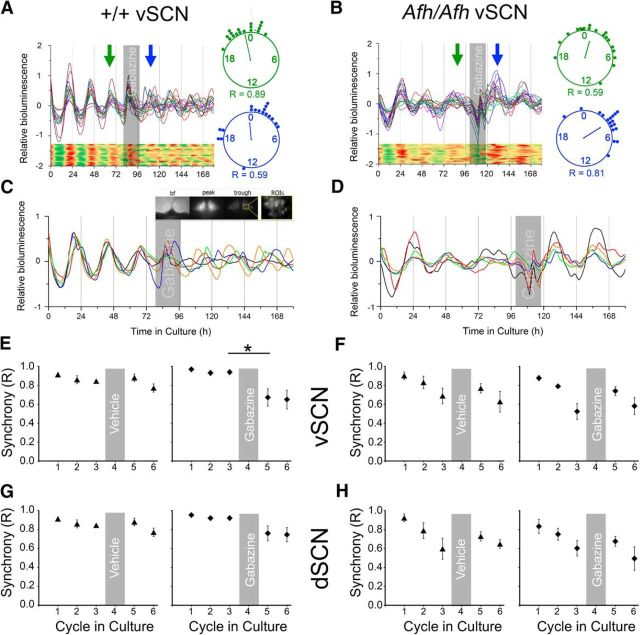
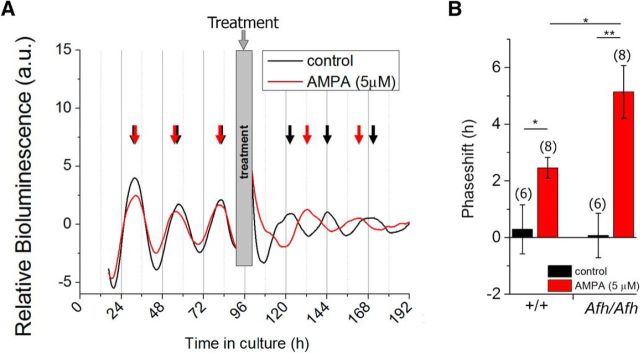
Suprachiasmatic nuclei (SCN) neurons contain an intracellular molecular circadian clock and the Cryptochromes (CRY1/2), key transcriptional repressors of this molecular apparatus, are subject to post-translational modification through ubiquitination and targeting for proteosomal degradation by the ubiquitin E3 ligase complex. Loss-of-function point mutations in a component of this ligase complex, Fbxl3, delay CRY1/2 degradation, reduce circadian rhythm strength, and lengthen the circadian period by ∼2.5 h. The molecular clock drives circadian changes in the membrane properties of SCN neurons, but it is unclear how alterations in CRY1/2 stability affect SCN neurophysiology. Here we use male and female Afterhours mice which carry the circadian period lengthening loss-of-function Fbxl3Afh mutation and perform patch-clamp recordings from SCN brain slices across the projected day/night cycle. We find that the daily rhythm in membrane excitability in the ventral SCN (vSCN) was enhanced in amplitude and delayed in timing in Fbxl3Afh/Afh mice. At night, vSCN cells from Fbxl3Afh/Afh mice were more hyperpolarized, receiving more GABAergic input than their Fbxl3+/+ counterparts. Unexpectedly, the progression to daytime hyperexcited states was slowed by Afh mutation, whereas the decline to hypoexcited states was accelerated. In long-term bioluminescence recordings, GABAA receptor blockade desynchronized the Fbxl3+/+ but not the Fbxl3Afh/Afh vSCN neuronal network. Further, a neurochemical mimic of the light input pathway evoked larger shifts in molecular clock rhythms in Fbxl3Afh/Afh compared with Fbxl3+/+ SCN slices. These results reveal unanticipated consequences of delaying CRY degradation, indicating that the Afh mutation prolongs nighttime hyperpolarized states of vSCN cells through increased GABAergic synaptic transmission.SIGNIFICANCE STATEMENT The intracellular molecular clock drives changes in SCN neuronal excitability, but it is unclear how mutations affecting post-translational modification of molecular clock proteins influence the temporal expression of SCN neuronal state or intercellular communication within the SCN network. Here we show for the first time, that a mutation that prolongs the stability of key components of the intracellular clock, the cryptochrome proteins, unexpectedly increases in the expression of hypoexcited neuronal state in the ventral SCN at night and enhances hyperpolarization of ventral SCN neurons at this time. This is accompanied by increased GABAergic signaling and by enhanced responsiveness to a neurochemical mimic of the light input pathway to the SCN. Therefore, post-translational modification shapes SCN neuronal state and network properties.

Keywords: brain slice; circadian; cryptochrome; electrophysiology; fbxl3; post-translational modification; suprachiasmatic.


Figures

Figure 1.
Figure 1.
Reduced excitability of Fbxl3Afh/Afh vSCN neurons. AD, Four different electrophysiological states of SCN neurons were initially distinguished in both Fbxl3+/+ (+/+) and Fbxl3Afh/Afh (Afh/Afh) brain slices: (A) depolarized silent, (B) depolarized with low-amplitude membrane oscillations or DLAMO, (C) spontaneously firing, and (D) hyperpolarized silent. Broken horizontal black line in AD delineates the resting membrane potential for the recording. E, Recordings were made with pipettes visually guided into either the dSCN or vSCN depicted in the illustration (Ee1) that corresponds to delineated white-outlined ovals in the photomicrograph (Ee2) of live coronal SCN brain slice. 3V, Third ventricle; OX, optic chiasm. Broken orange lines highlight a recording pipette targeted at dSCN. F, Percentages of cells in the different electrophysiological states (color coded as above, except cells in depolarized states A and B are collated (orange) for analysis as 1 category) during the day (top row) and night (bottom row) are shown as pie charts for +/+ and Afh/Afh genotypes. For the whole SCN, cell state was significantly associated with time of day for each genotype, with hyperpolarized states tending to be more numerous at night, particularly in the Afh/Afh SCN. G, The mean RMP of Fbxl3Afh/Afh vSCN neurons was more hyperpolarized than that of +/+ vSCN neurons during the early (ZT12–ZT18) and later (ZT18–ZT24) night. No genotype differences were detected for RMP in the dSCN (data not shown). Data in G are plotted as mean ± SEM. **p < 0.01, ***p < 0.001.
Figure 2.
Figure 2.
Fbxl3Afh mutation asymmetrically alters the daily rise and decline in membrane excitability of SCN neurons. The peak and nadir of resting membrane potential (A, B) and spontaneous firing rate (C) of vSCN neurons were delayed by the Afh mutation. In the vSCN of Fbxl3Afh/Afh (Afh/Afh) mice, the daytime progression from nadir to peak resting membrane potential slowed (∼1.4 h; A, B), whereas the decline to the nocturnal nadir was accelerated (∼1.4 h). At night, the RMP was more hyperpolarized than that of Fbxl3+/+ vSCN neurons (+/+), whereas spontaneous firing rate did not differ between the genotypes (C). Prominent asymmetry in the daily increase and decrease in RMP was seen in the FbxlAfh/Afh vSCN (B). Intergenotype comparison of the fitted spline with the GLIMMIX procedure indicated that RMP of Afh/Afh vSCN neurons differed from the RMP of +/+ SCN neurons across each ZT hour from ZT14–ZT22 (range p < 0.05–p < 0.001; A, B). Time of peak and nadir in firing rate were delayed by ∼2.0 and 0.9 h, respectively, in the Afh/Afh vSCN (C). Compared with the +/+ dSCN, time of maximal and minimal RMP were delayed by ∼1.3 and ∼2.8 h, respectively, in the Afh/Afh dSCN (D, E). However, no intergenotype differences were detected in RMP of dSCN neurons across the projected day/night cycle (D, E). The asymmetry in the daily increase and decrease in RMP seen in the Fbxl3+/+ dSCN was reversed in the dSCN of Fbxl3Afh/Afh mice (E). In the dSCN, the amplitude of the firing rate rhythm was reduced in the Fbxl3Afh/Afh compared with Fbxl3+/+ animals (0.4 vs 1.4 Hz), whereas the time of peak and nadir in firing rate of dSCN neurons showed very small genotype-related differences (F). Data in A and D are scatter plots of individual data points (Afh/Afh in red unfilled circles; +/+ in blue unfilled circles) fitted with cubic spline fits of the mean ± 95% confidence interval, whereas data in B, C, E, and F are plotted as cubic spline fits of the mean ± 95% confidence interval. B and E are the cubic splines in A and D, respectively, plotted against an expanded ordinate.
Figure 3.
Figure 3.
The hyperpolarized state of Fbxl3Afh/Afh vSCN neurons was sustained during the circadian night. A, Examples of wheel-running activity (WRA) rhythms of Fbxl3+/+ (+/+) and Fbxl3Afh/Afh (Afh/Afh) mice in constant dark. WRA: CT12 = activity onset (black arrows). Circadian times of recorded cells were adjusted according to the circadian period of individual animals as calculated from activity onsets. The circadian period in WRA was lengthened by the Afh mutation (B), whereas rhythm strength was diminished (C). During circadian night, RMP was reduced by the Afh mutation (D), whereas spontaneous firing rate was slightly damped (E). Data in BE are plotted as mean ± SEM. *p < 0.05, ***p < 0.001.
Figure 4.
Figure 4.
The Fbxl3Afh mutation elevates the frequency of GABAergic synaptic events in vSCN neurons during the night. In voltage-clamp recordings made at a holding potential of −70 mV, spontaneous synaptic events present in SCN slices from Fbxl3+/+ (+/+) and Fbxl3Afh/Afh (Afh/Afh) mice (A, top traces) were unaffected by glutamatergic antagonists (CNQX blocks AMPA receptors, AP-5 blocks N-methyl-D-aspartate (NMDA) receptors; A, middle traces), but abolished by the addition of the GABA antagonist, gabazine (A, bottom traces). This indicates that GABAergic and not glutamatergic sPSCs, are predominant in the SCN of Fbxl3+/+ and Fbxl3Afh/Afh animals. B, Compared with the night, the frequency of the GABAergic sPSCs was significantly higher during the day in the dSCN but not the vSCN of Fbxl3+/+ mice. By contrast, in the vSCN of Fbxl3Afh/Afh mice, the night-time frequency of the GABAergic sPSCs was significantly higher than that observed during the day, and significantly higher than that recorded in the vSCN of Fbxl3+/+ mice at this time. Further the daytime frequency of GABAergic sPSCs in the dSCN of Fbxl3Afh/Afh animals was significantly lower than that recorded during the day in the dSCN of Fbxl3+/+ mice. Data in B are plotted as mean ± SEM. Within-genotype differences are denoted by **p < 0.01, and between-genotype differences denoted by #p < 0.05, ##p < 0.01.
Figure 5.
Figure 5.
Blockade of GABA signaling fails to desynchronize PER2::LUC single-cell rhythms in the Fbxl3Afh/Afh SCN. PER2::LUC single-cell bioluminescence recordings discriminated in Fbxl3+/+ (+/+; A, C, E, G) and Fbxl3Afh/Afh (Afh/Afh; B, D, F, H) SCN slice preparations. From each slice 18–22 cell-like ROIs were discriminated from the dSCN and vSCN subregions. Synchrony (R) was assessed before (cycles 1–3) and following treatment (cycles 5–6), and single-cell heatmaps of the representative traces are shown for better visualization (A and B show data from vSCN). Gray shaded areas depict time of treatment. Rayleigh plots and R values are shown for cycles 3 (green) and 4 (blue) of the representative traces of Fbxl3+/+ (A) and Fbxl3Afh/Afh (B) genotypes, with the clock hands depicting the mean peak time and their length representing the R value (the longer the clock hand, the larger the R value). C, D, Recordings of five cells from a vSCN slice of each genotype are shown in magnification and example bf, peak, and trough bioluminescence images from Fbxl3+/+ vSCN are depicted in C (inset). Gabazine was detrimental to synchrony in the Fbxl3+/+ vSCN, whereas vehicle treatment was not (C, E). Neither vehicle nor gabazine had significant effects on cell synchrony in the Fbxl3+/+ dSCN (G). The desynchronizing action of gabazine was not detected in the Fbxl3Afh/Afh vSCN and dSCN (D, F, H). Vehicle treatment of the SCN was without effect on synchrony in either vSCN (F) or dSCN (H) of the Fbxl3Afh/Afh SCN tissue slices. Data in EH are plotted as mean ± SEM. *p < 0.05.
Figure 6.
Figure 6.
Phase-advancing effects of AMPA are enhanced in the Fbxl3Afh/Afh SCN. A, Example traces of photomultiplier tube bioluminescence recordings from an Fbxl3Afh/Afh (Afh/Afh) SCN explant treated with vehicle (black trace) or 5 μm AMPA (red trace) at the trough of day 5 in culture. The arrows indicate the point of peak expression used for calculation of the period, with red arrows delineating peaks pre- and post-AMPA treatment and black arrows showing peaks prevehicle and postvehicle control treatment. B, Compared with vehicle treatment, AMPA evoked significant phase advance in the timing of peak PER2::LUC bioluminescence in both Fbxl3+/+ (+/+) and Fbxl3Afh/Afh SCN explants. Larger advances were evoked by AMPA in Fbxl3Afh/Afh compared with Fbxl3+/+ SCN explants (5.1 ± 0.9 vs 2.5 ± 0.4 h, p = 0.0018), sample sizes given in brackets. *p < 0.05, **p < 0.01.

Similar articles

Cited by

References

    1. Albers HE, Walton JC, Gamble KL, McNeill JK 4th, Hummer DL (2017) The dynamics of GABA signaling: revelations from the circadian pacemaker in the suprachiasmatic nucleus. Front Neuroendocrinol 44:35–82. 10.1016/j.yfrne.2016.11.003 - DOI - PMC - PubMed
    1. Albus H, Vansteensel MJ, Michel S, Block GD, Meijer JH (2005) A GABAergic mechanism is necessary for coupling dissociable ventral and dorsal regional oscillators within the circadian clock. Curr Biol 15:886–893. 10.1016/j.cub.2005.03.051 - DOI - PubMed
    1. Anand SN, Maywood ES, Chesham JE, Joynson G, Banks GT, Hastings MH, Nolan PM (2013) Distinct and separable roles for endogenous CRY1 and CRY2 within the circadian molecular clockwork of the suprachiasmatic nucleus, as revealed by the Fbxl3(Afh) mutation. J Neurosci 33:7145–7153. 10.1523/JNEUROSCI.4950-12.2013 - DOI - PMC - PubMed
    1. Antle MC, Silver R (2005) Orchestrating time: arrangements of the brain circadian clock. Trends Neurosci 28:145–151. 10.1016/j.tins.2005.01.003 - DOI - PubMed
    1. Azzi A, Dallmann R, Casserly A, Rehrauer H, Patrignani A, Maier B, Kramer A, Brown SA (2014) Circadian behavior is light-reprogrammed by plastic DNA methylation. Nat Neurosci 17:377–382. 10.1038/nn.3651 - DOI - PubMed

Publication types

LinkOut - more resources