Review
. 2017 Jul 9;6(3):21.
doi: 10.3390/cells6030021.

Studying Autophagy in Zebrafish

Affiliations
Review

Studying Autophagy in Zebrafish

Benan John Mathai et al. Cells. .

Abstract

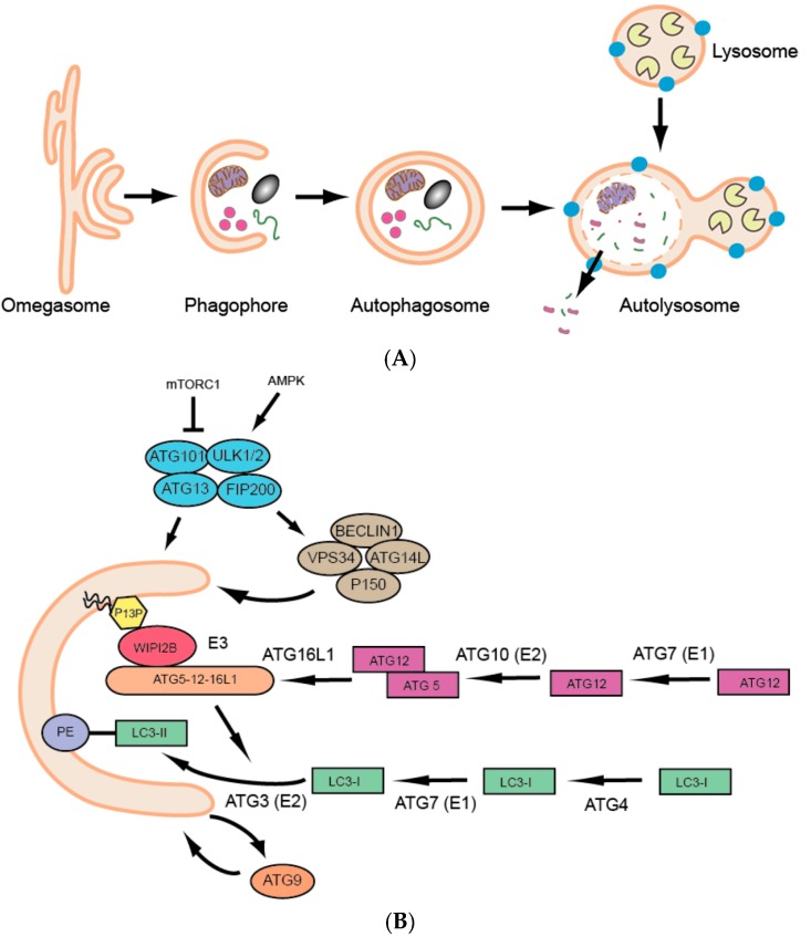
Autophagy is an evolutionarily conserved catabolic process which allows lysosomal degradation of complex cytoplasmic components into basic biomolecules that are recycled for further cellular use. Autophagy is critical for cellular homeostasis and for degradation of misfolded proteins and damaged organelles as well as intracellular pathogens. The role of autophagy in protection against age-related diseases and a plethora of other diseases is now coming to light; assisted by several divergent eukaryotic model systems ranging from yeast to mice. We here give an overview of different methods used to analyse autophagy in zebrafish-a relatively new model for studying autophagy-and briefly discuss what has been done so far and possible future directions.

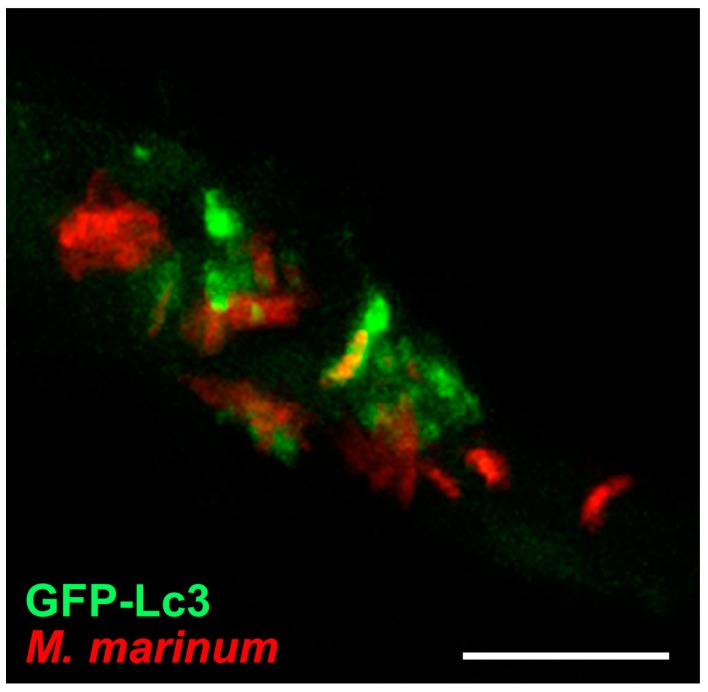
Keywords: GFP-Lc3; aggrephagy; autophagy; confocal microscopy; mitophagy; xenophagy; zebrafish.


Conflict of interest statement

The authors have no conflicts of interest.

Figures

Figure 1
Figure 1
(A) Schematic overview of the process of macroautophagy; (B) Schematic overview of the core autophagic proteins involved in autophagosome biogenesis.
Figure 2
Figure 2
Messenger RNA sequence (mRNA-seq) analysis. Line plot of core autophagy gene transcripts analyzed by mRNA-seq in zebrafish embryos from the oocyte stage to post-mid blastula stage transition.
Figure 3
Figure 3
Confocal imaging of Tg(CMV:GFP-Lc3). Representative confocal images of GFP-Lc3 puncta (autophagosomes) in the trunk area of GFP-Lc3 transgenic zebrafish embryos injected with control morpholino or Hslbp3 translational-blocking morpholino and imaged at 2 days post fertilization (dpf) with or without pre-treatment with chloroquine (10 mM) for 6 h. Scale bars, 10 µM for the confocal images. Panel A, B shows the whole zebrafish larvae at 2 days post fertilization highlighting the trunk area chosen for confocal imaging; Panel A’, A’’, B’, B’’ shows respective confocal images.
Figure 4
Figure 4
Schematic overview of the autophagic pathway and a partial list of reagents (reagents used beyond 2014, Table 2) that modulate autophagy in zebrafish are indicated.
Figure 5
Figure 5
Confocal imaging of Tg(CMV:EGFP-Mapllc3b) on infection. GFP-Lc3 signal around clusters of M. marinum bacteria in 4-day-old zebrafish larva at 3 days post infection. Scale bars, 10 µM.

References

    1. Boya P., Reggiori F., Codogno P. Emerging regulation and functions of autophagy. Nat. Cell Biol. 2013;15:713–720. doi: 10.1038/ncb2788. - DOI - PMC - PubMed
    1. Takeshige K., Baba M., Tsuboi S., Noda T., Ohsumi Y. Autophagy in Yeast Demonstrated with Proteinase-deficient Mutants and Conditions for its Induction. J. Cell Biol. 1992;119:301–311. doi: 10.1083/jcb.119.2.301. - DOI - PMC - PubMed
    1. Tsukada M., Ohsumi Y. Isofation and charactization of autophagy-defective mutants of Saccharomyces cerevisiae. FEBS Lett. 1993;333:169–174. doi: 10.1016/0014-5793(93)80398-E. - DOI - PubMed
    1. Thumm M., Egner R., Koch B., Schlumpberger M., Straub M., Veenhuis M., Wolf D.H. Isolation of autophagocytosis mutants of Saccharomyces cerevisiae. FEBS Lett. 1994;349:275–280. doi: 10.1016/0014-5793(94)00672-5. - DOI - PubMed
    1. Harding T.M., Morano K.A., Scott S.V., Klionsky D.J. Isolation and characterization of yeast mutants in the cytoplasm to vacuole protein targeting pathway. J. Cell Biol. 1995;131:591–602. doi: 10.1083/jcb.131.3.591. - DOI - PMC - PubMed