CD15s/CD62E Interaction Mediates the Adhesion of Non-Small Cell Lung Cancer Cells on Brain Endothelial Cells: Implications for Cerebral Metastasis
- PMID: 28698503
- PMCID: PMC5535965
- DOI: 10.3390/ijms18071474
CD15s/CD62E Interaction Mediates the Adhesion of Non-Small Cell Lung Cancer Cells on Brain Endothelial Cells: Implications for Cerebral Metastasis
Abstract
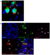
Expression of the cell adhesion molecule (CAM), Sialyl Lewis X (CD15s) correlates with cancer metastasis, while expression of E-selectin (CD62E) is stimulated by TNF-α. CD15s/CD62E interaction plays a key role in the homing process of circulating leukocytes. We investigated the heterophilic interaction of CD15s and CD62E in brain metastasis-related cancer cell adhesion. CD15s and CD62E were characterised in human brain endothelium (hCMEC/D3), primary non-small cell lung cancer (NSCLC) (COR-L105 and A549) and metastatic NSCLC (SEBTA-001 and NCI-H1299) using immunocytochemistry, Western blotting, flow cytometry and immunohistochemistry in human brain tissue sections. TNF-α (25 pg/mL) stimulated extracellular expression of CD62E while adhesion assays, under both static and physiological flow live-cell conditions, explored the effect of CD15s-mAb immunoblocking on adhesion of cancer cell-brain endothelium. CD15s was faintly expressed on hCMEC/D3, while high levels were observed on primary NSCLC cells with expression highest on metastatic NSCLC cells (p < 0.001). CD62E was highly expressed on hCMEC/D3 cells activated with TNF-α, with lower levels on primary and metastatic NSCLC cells. CD15s and CD62E were expressed on lung metastatic brain biopsies. CD15s/CD62E interaction was localised at adhesion sites of cancer cell-brain endothelium. CD15s immunoblocking significantly decreased cancer cell adhesion to brain endothelium under static and shear stress conditions (p < 0.001), highlighting the role of CD15s-CD62E interaction in brain metastasis.
Keywords: CD15s; CD62E; Sialyl Lewis X; adhesion; brain metastasis.
Conflict of interest statement
The authors declare no conflict of interest.
Figures


Similar articles
-
Carcinoembryonic antigen is a sialyl Lewis x/a carrier and an E‑selectin ligand in non‑small cell lung cancer.Int J Oncol. 2019 Nov;55(5):1033-1048. doi: 10.3892/ijo.2019.4886. Epub 2019 Sep 26. Int J Oncol. 2019. PMID: 31793656 Free PMC article.
-
TNF-α enhancement of CD62E mediates adhesion of non-small cell lung cancer cells to brain endothelium via CD15 in lung-brain metastasis.Neuro Oncol. 2016 May;18(5):679-90. doi: 10.1093/neuonc/nov248. Epub 2015 Oct 15. Neuro Oncol. 2016. PMID: 26472821 Free PMC article.
-
Fucosyltransferase 4 and 7 mediates adhesion of non-small cell lung cancer cells to brain-derived endothelial cells and results in modification of the blood-brain-barrier: in vitro investigation of CD15 and CD15s in lung-to-brain metastasis.J Neurooncol. 2019 Jul;143(3):405-415. doi: 10.1007/s11060-019-03188-x. Epub 2019 May 18. J Neurooncol. 2019. PMID: 31104223 Free PMC article.
-
SA-Lea and tumor metastasis: the old prediction and recent findings.Hybrid Hybridomics. 2002 Apr;21(2):111-6. doi: 10.1089/153685902317401708. Hybrid Hybridomics. 2002. PMID: 12031100 Review.
-
Carbohydrate-mediated cell adhesion involved in hematogenous metastasis of cancer.Glycoconj J. 1997 Aug;14(5):577-84. doi: 10.1023/a:1018532409041. Glycoconj J. 1997. PMID: 9298690 Review.
Cited by
-
Inflammatory Environment Promotes the Adhesion of Tumor Cells to Brain Microvascular Endothelial Cells.Front Oncol. 2021 Jun 16;11:691771. doi: 10.3389/fonc.2021.691771. eCollection 2021. Front Oncol. 2021. PMID: 34222020 Free PMC article.
-
Insights into the Molecular Mechanisms Mediating Extravasation in Brain Metastasis of Breast Cancer, Melanoma, and Lung Cancer.Cancers (Basel). 2023 Apr 12;15(8):2258. doi: 10.3390/cancers15082258. Cancers (Basel). 2023. PMID: 37190188 Free PMC article. Review.
-
A novel molecular magnetic resonance imaging agent targeting activated leukocyte cell adhesion molecule as demonstrated in mouse brain metastasis models.J Cereb Blood Flow Metab. 2021 Jul;41(7):1592-1607. doi: 10.1177/0271678X20968943. Epub 2020 Nov 5. J Cereb Blood Flow Metab. 2021. PMID: 33153376 Free PMC article.
-
Carcinoembryonic antigen is a sialyl Lewis x/a carrier and an E‑selectin ligand in non‑small cell lung cancer.Int J Oncol. 2019 Nov;55(5):1033-1048. doi: 10.3892/ijo.2019.4886. Epub 2019 Sep 26. Int J Oncol. 2019. PMID: 31793656 Free PMC article.
-
Cardiac Dysfunction Promotes Cancer Progression via Multiple Secreted Factors.Cancer Res. 2022 May 3;82(9):1753-1761. doi: 10.1158/0008-5472.CAN-21-2463. Cancer Res. 2022. PMID: 35260887 Free PMC article.
References
-
- Narimatsu H., Iwasaki H., Nishihara S., Kimura H., Kudo T., Yamauchi Y., Hirohashi S. Genetic evidence for the Lewis enzyme, which synthesizes type-1 Lewis antigens in colon tissue and intracellular localization of the enzyme. Cancer Res. 1996;56:330–338. - PubMed
-
- Martin K., Akinwunmi J., Rooprai H.K., Kennedy A.J., Linke A., Ognjenovic N., Pilkington G.J. Non expression of CD15 by neoplastic glia: A barrier to metastasis? Anticancer Res. 1995;15:1159–1166. - PubMed
MeSH terms
Substances
LinkOut - more resources
Full Text Sources
Other Literature Sources
Medical

