Review
. 2017 Jul 10;18(7):1487.
doi: 10.3390/ijms18071487.

Altered Mitochondrial Metabolism and Mechanosensation in the Failing Heart: Focus on Intracellular Calcium Signaling

Affiliations
Review

Altered Mitochondrial Metabolism and Mechanosensation in the Failing Heart: Focus on Intracellular Calcium Signaling

Aderville Cabassi et al. Int J Mol Sci. .

Abstract

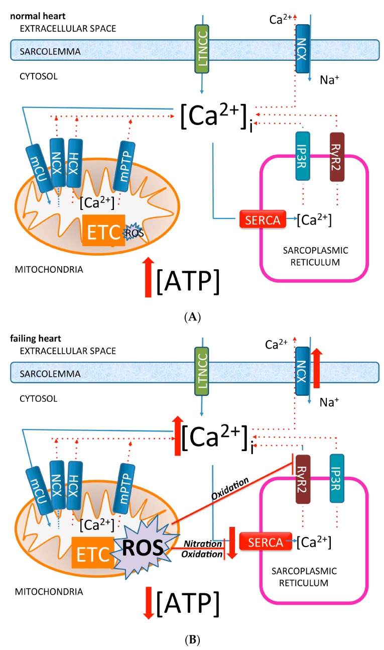
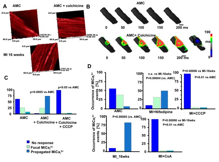
The heart consists of millions of cells, namely cardiomyocytes, which are highly organized in terms of structure and function, at both macroscale and microscale levels. Such meticulous organization is imperative for assuring the physiological pump-function of the heart. One of the key players for the electrical and mechanical synchronization and contraction is the calcium ion via the well-known calcium-induced calcium release process. In cardiovascular diseases, the structural organization is lost, resulting in morphological, electrical, and metabolic remodeling owing the imbalance of the calcium handling and promoting heart failure and arrhythmias. Recently, attention has been focused on the role of mitochondria, which seem to jeopardize these events by misbalancing the calcium processes. In this review, we highlight our recent findings, especially the role of mitochondria (dys)function in failing cardiomyocytes with respect to the calcium machinery.

Keywords: calcium transient; fatty acids; mechanoelectric transduction; microdomains; mitochondria metabolism.


Conflict of interest statement

The authors declare no conflict of interest.

Figures

Figure 1
Figure 1
Diagram of subcellular interaction between subcellular calcium compartmentation and reactive oxygen species (ROS) in normal (A) and failing (B) cardiomyocytes. LTCC: L-Type Calcium Channel; NCX: Sodium-Calcium Exchanger; HCX: Hydrogen-Calcium Exchanger; mCU: Mitochondria Calcium Uniporter; mPTP: Mitochondrial Permeability Transition Pore; SERCA: Sarco/Endoplasmic Reticulum Ca2+-ATPase; IP3R: Inositol Trisphosphate Receptors; RyR: Ryanodine Receptors; ROS: Reactive Oxygen Species; ETC: Electron Transport Chain. Dotted lines indicate Ca2+ efflux; full line indicate Ca2+ influx.
Figure 2
Figure 2
Schematic representation of nanoscopic technology for interrogates subsarcolemmal mitochondrial mechanosensitivty. Blue dotted line: excitation light. Green dotted line: emission light from the sample.
Figure 3
Figure 3
Membrane compliance related to mechanically induced Ca2+ release. (A) Left. SICM topographical image of a failing rat ventricular cardiomyocyte. Asterisks indicate the position where hydrojet pressures were applied (black = partially striated area, red = unstriated area). Right. Pressure vs. Z displacement in the two positions selected on SICM image. To note the unstriated area is stiffer; (B) Membrane compliance (µm/kPa) during progression toward HF (MI-16 weeks (wks)) in crest, groove, and unstriated areas; (C) Mechanically-induced Ca2+ release after hydrojet pressure delivery, in AMC cell top and in HF cell bottom. * p < 0.05 vs. control; ** p < 0.001 vs. control. Modified from [6] with permission.
Figure 4
Figure 4
Surface scanning ion conductance microscopy analysis of mitochondrial displacement. (A) SICM images of 10 μm × 10 μm cardiomyocyte regions of AMC (top) and MI (bottom) cells; (B) Surface confocal images (obtained by SSICM) of the labelled TMRM active mitochondria in (A); (C) Merged images for SICM topography TMRM-labelled mitochondria; (D) Mitochondrial displacement observed by TEM. Scale bar: 500 nm Modified from [6] with permission.
Figure 5
Figure 5
Microtubule network derangements together with mitochondrial displacement are prerequisite for mechanically induced Ca2+ release initiation. (A) TMRM-labelled mitochondria position in AMC, AMC+colchicine, and HF (MI-16 weeks); (B) Mechanically induced Ca2+ initiation in AMC cell (top) and the same cell in the presence of colchicine (bottom); (C) Occurrence of MiCai events in terms of no response, focal MiCai and total MiCai in AMC, AMC+colchicine, AMC+colchcine+CCCP; (D) Same as C for AMC, MI-16 wks, MI-16-wks + Nifedipine, MI-16-wks + CCCP, MI-16-wks + CsA. Modified from [6] with permission.

Similar articles

Cited by

References

    1. Iribe G., Ward C.W., Camelliti P., Bollensdorff C., Mason F., Burton R.A., Garny A., Morphew M.K., Hoenger A., Lederer W.J., et al. Axial stretch of rat single ventricular cardiomyocytes causes an acute and transient increase in Ca2+ spark rate. Circ. Res. 2009;104:787–795. doi: 10.1161/CIRCRESAHA.108.193334. - DOI - PMC - PubMed
    1. Link M.S., Wang P.J., Pandian N.G., Bharati S., Udelson J.E., Lee M.Y., Vecchiotti M.A., VanderBrink B.A., Mirra G., Maron B.J., et al. An experimental model of sudden death due to low-energy chest-wall impact (commotio cordis) N. Engl. J. Med. 1998;338:1805–1811. doi: 10.1056/NEJM199806183382504. - DOI - PubMed
    1. Kiseleva I., Kamkin A., Wagner K.D., Theres H., Ladhoff A., Scholz H., Gunther J., Lab M.J. Mechanoelectric feedback after left ventricular infarction in rats. Cardiovasc. Res. 2000;45:370–378. doi: 10.1016/S0008-6363(99)00361-2. - DOI - PubMed
    1. Lammerding J., Kamm R.D., Lee R.T. Mechanotransduction in cardiac myocytes. Ann. N. Y. Acad. Sci. 2004;1015:53–70. doi: 10.1196/annals.1302.005. - DOI - PubMed
    1. Kohlhaas M., Maack C. Calcium release microdomains and mitochondria. Cardiovasc. Res. 2013;98:259–268. doi: 10.1093/cvr/cvt032. - DOI - PubMed

LinkOut - more resources