. 2017 Jul 10;17(7):1603.
doi: 10.3390/s17071603.

Microfluidic Platform for the Long-Term On-Chip Cultivation of Mammalian Cells for Lab-On-A-Chip Applications

Affiliations

Microfluidic Platform for the Long-Term On-Chip Cultivation of Mammalian Cells for Lab-On-A-Chip Applications

Frank Bunge et al. Sensors (Basel). .

Abstract

Lab-on-a-Chip (LoC) applications for the long-term analysis of mammalian cells are still very rare due to the lack of convenient cell cultivation devices. The difficulties are the integration of suitable supply structures, the need of expensive equipment like an incubator and sophisticated pumps as well as the choice of material. The presented device is made out of hard, but non-cytotoxic materials (silicon and glass) and contains two vertical arranged membranes out of hydrogel. The porous membranes are used to separate the culture chamber from two supply channels for gases and nutrients. The cells are fed continuously by diffusion through the membranes without the need of an incubator and low requirements on the supply of medium to the assembly. The diffusion of oxygen is modelled in order to find the optimal dimensions of the chamber. The chip is connected via 3D-printed holders to the macroscopic world. The holders are coated with Parlyene C to ensure that only biocompatible materials are in contact with the culture medium. The experiments with MDCK-cells show the successful seeding inside the chip, culturing and passaging. Consequently, the presented platform is a step towards Lab-on-a-Chip applications that require long-term cultivation of mammalian cells.

Keywords: Lab-on-a-Chip; MDCK; cell cultivation; diffusion model; hydrogel; parylene.


Conflict of interest statement

The authors declare no conflict of interest. The founding sponsors had no role in the design of the study; in the collection, analyses, or interpretation of data; in the writing of the manuscript, and in the decision to publish the results.

Figures

Figure 1
Figure 1
Concept of the microfluidic chip for the long-term cultivation of mammalian cells in a lab-on-a-chip context as a cross-section. The half of the chip that is cut away for better visualisation is identical to the shown one. The figure is not to scale.
Figure 2
Figure 2
Course of the concentration of oxygen c(O2) in the steady state is shown for wHg=1.1 mm, h=0.38 mm, Ac=400 µm2, D=2.4×10⁻⁹ m2s, a width of the culture chamber of 5 mm and different filling factors γ.
Figure 3
Figure 3
Maximal width of the growth chamber wGr based on the analytical model: (a) width of the chamber as a function of the chamber height h for different filling factors γ and different α describing the ratio of the hydrogel width to the chamber height; (b) width of the chamber as a function of the filling factor γ, which describes the ratio of the surface that is covered with cells for different heights and α.
Figure 4
Figure 4
Fabrication of the device: (1) Deep reactive ion etching (DRIE) process for the channels into 380 µm thick silicon; (2) patterning of titanium nitride and gold on two 520 µm thick borosilicateglass wafers; (3) anodic bonding of one glass wafer and the silicon wafer; (4) powderblasting of the inlets into the second glass wafer; (5) anodic bonding; (6) coating of gold with octadecanethiol (ODT); (7) creation of the agarose membranes.
Figure 5
Figure 5
Image of the fabricated chip. The size of the chip is 13 × 17 × 1.4 mm3.
Figure 6
Figure 6
Assembly of the microfluidic chip that is clamped between 3D-printed holders and sealed with O-rings: (a) model of the assembly showing all components (b) image of the assembly with the dimensions of 29 × 21.7 × 7.9 mm.
Figure 7
Figure 7
Culture of MDCK-cells with GMEM-medium 24 h after seeding: (a) negative sample without any 3D-printed parts showing high cell viability; (b) cell culture in which a 3D-printed part out of HTM140 is inserted showing influence of the toxicity of the material; (c) cell culture with a 3D-printed part out of HTM140 that is coated with 10 µm Parylene C showing the same cell viability as the negative sample. Scale bar is 100 µm.
Figure 8
Figure 8
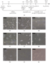
Culture of MDCK-cells (scale bar is 100 µm): (a) overview about the experimental procedure (not to scale); (b) t=0 h: seeding of the cells inside the chip; (c) t=4 h: cells adhere on the bottom plate; (d) t=24 h: cell growth inside the chamber; (e) t=24 h (culture) +12min incubation with TrypLE: first cells detach from the plate; (f) t=24 h+30min incubation with TrypLE: Detaching of almost all cells; (g) t=24.5 h+45min: A few cells remain inside the chip after the splitting; (h) t=24.5 h+24 h: on-chip growth of the cells; (i) t=24.5 h+45min: the majority of the cells is removed from the chip after splitting and added in a 24-well plate; (j) t=24.5 h+24 h: off-chip growth of the cells.

Similar articles

Cited by

References

    1. Chin C.D., Linder V., Sia S.K. Commercialization of microfluidic point-of-care diagnostic devices. Lab Chip. 2012;12:2118. doi: 10.1039/c2lc21204h. - DOI - PubMed
    1. Booth R., Kim H. Characterization of a microfluidic in vitro model of the blood-brain barrier. Lab Chip. 2012;12:1784. doi: 10.1039/c2lc40094d. - DOI - PubMed
    1. Schurink B., Luttge R. Hydrogel/poly-dimethylsiloxane hybrid bioreactor facilitating 3D cell culturing. J. Vac. Sci. Technol. B Nanotechnol. Microelectron. Mater. Process. Meas. Phenom. 2013;31:06F903. doi: 10.1116/1.4831762. - DOI
    1. Grist S.M., Chrostowski L., Cheung K.C. Optical Oxygen Sensors for Applications in Microfluidic Cell Culture. Sensors. 2010;10:9286–9316. doi: 10.3390/s101009286. - DOI - PMC - PubMed
    1. Yu T., Guo Z., Fan H., Song J., Liu Y., Gao Z., Wang Q. Cancer-associated fibroblasts promote non-small cell lung cancer cell invasion by upregulation of glucose-regulated protein 78 (GRP78) expression in an integrated bionic microfluidic device. Oncotarget. 2016;7:25593. doi: 10.18632/oncotarget.8232. - DOI - PMC - PubMed

Substances

LinkOut - more resources