. 2017 Jul 11;7(1):5152.
doi: 10.1038/s41598-017-04916-x.

Laminin-511 and -521-based matrices for efficient ex vivo-expansion of human limbal epithelial progenitor cells

Affiliations

Laminin-511 and -521-based matrices for efficient ex vivo-expansion of human limbal epithelial progenitor cells

Naresh Polisetti et al. Sci Rep. .

Abstract

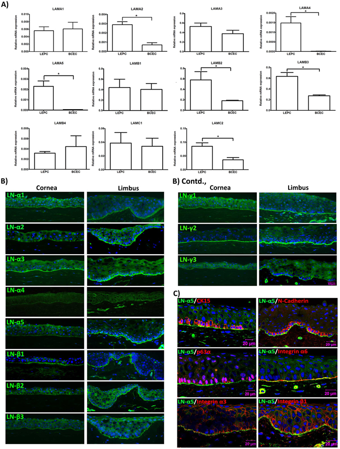
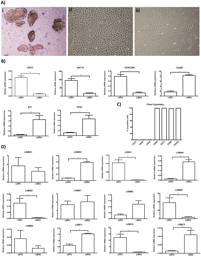
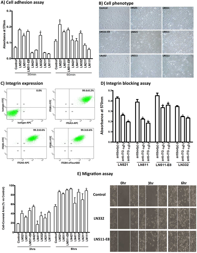
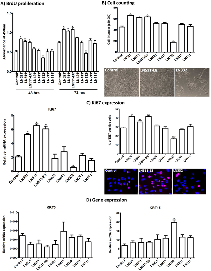
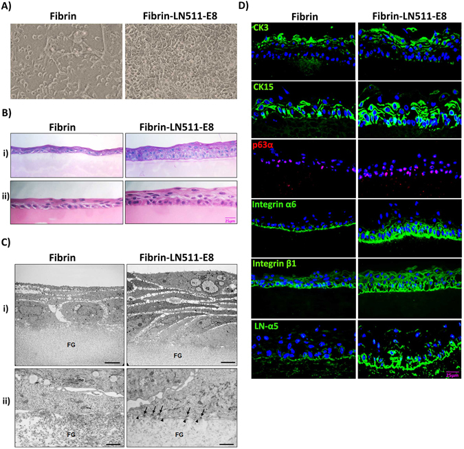
Optimization of culture conditions for human limbal epithelial stem/progenitor cells (LEPC) that incorporate the in vivo cell-matrix interactions are essential to enhance LEPC ex vivo-expansion and transplantation efficiency. Here, we investigate the efficacy of laminin (LN) isoforms preferentially expressed in the limbal niche as culture matrices for epithelial tissue engineering. Analyses of expression patterns of LN chains in the human limbal niche provided evidence for enrichment of LN-α2, -α3, -α5, -β1, -β2, -β3, -γ1, -γ2 and -γ3 chains in the limbal basement membrane, with LN-α5 representing a signature component specifically produced by epithelial progenitor cells. Recombinant human LN-521 and LN-511 significantly enhanced in vitro LEPC adhesion, migration and proliferation compared to other isoforms, and maintained phenotype stability. The bioactive LN-511-E8 fragment carrying only C-terminal domains showed similar efficacy as full-length LN-511. Functional blocking of α3β1 and α6β1 integrins suppressed adhesion of LEPC to LN-511/521-coated surfaces. Cultivation of LEPC on fibrin-based hydrogels incorporating LN-511-E8 resulted in firm integrin-mediated adhesion to the scaffold and well-stratified epithelial constructs, with maintenance of a progenitor cell phenotype in their (supra)basal layers. Thus, the incorporation of chemically defined LN-511-E8 into biosynthetic scaffolds represents a promising approach for xeno-free corneal epithelial tissue engineering for ocular surface reconstruction.


Conflict of interest statement

The authors declare that they have no competing interests.

Figures

Figure 1
Figure 1
Expression of laminin chains in the limbal stem cell niche in situ. (A) Quantitative real-time polymerase chain reaction (qRT-PCR) primer assays showing higher expression levels of laminin α2 (LAMA2), α4 (LAMA4), α5 (LAMA5), β2 (LAMB2), β3 (LAMB3), and γ2 (LAMC2) in microdissected limbal epithelial stem/progenitor cell (LEPC) clusters compared with basal corneal epithelial cell (BCEC) populations; laminin α1 (LAMA1), α3 (LAMA3), β1 (LAMB1), β4 (LAMB4), γ1 (LAMC1) showed no differential expression patterns. Data are expressed as means (2−ΔCT × 1,000) ± SEM (n = 5); *p < 0.05; Mann-Whitney U test. (B) Immunofluorescence analyses of corneoscleral tissue sections showing differential staining patterns of laminin α2, α5, β2, β3, γ2, and γ3, but similar staining patterns of laminin α1, α3, β1, and γ1 in the basement membranes of corneal and limbal epithelia; laminin α4 was largely negative in epithelial basement membranes. Nuclei are counterstained with DAPI (blue); scale bar = 60 µm. (C) Immunofluorescence double labeling of laminin (LN) α5 (green) and cytokeration (CK)15, N-Cadherin, p63α, integrin α6, integrin α3, and integrin β1 (red); nuclear counterstaining with DAPI (blue); scale bar = 20 µm.
Figure 2
Figure 2
Expression of laminin chains in limbal stem/progenitor and associated niche cells in vitro. (A) Phase contrast images of (i) limbal epithelial cell clusters after collagenase digestion, (ii) cultured limbal epithelial progenitor cells, and (iii) associated mesenchymal stromal cells. (B) Quantitative real-time polymerase chain reaction (qRT-PCR) primer assays confirming differential expression of established epithelial (KRT3, KRT15, CEACAM1) and mesenchymal (ICAM1, KIT, SOX2) markers in cultured limbal epithelial progenitor cells (LEPC) compared with cultured limbal mesenchymal stromal cells (LMSC). Data are expressed as means (2−ΔCT × 1,000) ± SEM (n = 5). (C) Flow cytometry analyses of cultured LMSC showing positive expression of CD44, CD73, CD90, and CD105, but negative expression of CD31, CD34, and CD45. Percentages (%) of positive cells are expressed as means ± SEM (n = 3). (D) qRT-PCR primer assays showing higher expression levels of laminin α3 (LAMA3), α5 (LAMA5), β3 (LAMB3), γ2 (LAMC2) in cultured LEPC, and higher expression levels of laminin α2 (LAMA2), α4 (LAMA4), β2 (LAMB2), γ1 (LAMC1), γ3 (LAMC3) in cultured LMSC; laminin α1 (LAMA1), β1 (LAMB1), and β4 (LAMB4) showed no differential expression patterns. Data are expressed as means (2−ΔCT × 1,000) ± SEM (n = 5); *p < 0.05; Mann-Whitney U test. (Abbreviations: KRT, Keratin; CEACAM1, carcinoembryonic antigen-related cell adhesion molecule 1; ICAM1, intercellular cell adhesion molecule 1; Sox2, sex determining region Y-box 2; CD, cluster of differentiation).
Figure 3
Figure 3
Effect of laminin isoforms on limbal progenitor cell adhesion and migration. (A) The effect of laminin (LN) isoforms on cell adhesion was tested by seeding limbal epithelial progenitor cells (LEPC) at a density of 50,000 cells/cm2 and spectrophotometric measurement of adherent cells 30 and 60 minutes after seeding. Data are expressed as means ± SEM (n = 4). (B) Phase contrast images of LEPC cultured on LN isoforms; magnification ×100. (C) Flow cytometry analyses of cultured LEPC showing expression of integrin α3 (ITGA3), integrin α6 (ITGA6), integrin β1 (ITGB1), and integrin β4 (ITGB4) on their surface. Percentages (%) of positive cells are expressed as means ± SEM percentage (%) (n = 3). (D) Functional blocking of integrin-mediated LEPC adhesion to LN-521, -511, -511-E8 and -332 was tested using neutralizing antibodies against integrin α3β1 and α6β1 60 minutes seeding. Data are expressed as means ± SEM (n = 4). (E) The effect of LN isoforms on LEPC migration was analyzed in two well-culture inserts with a defined cell-free gap and measurement of gap closure 3 and 6 hours after removal of the culture inserts. Data are expressed as means ± SEM (n = 3); *p < 0.05; Mann-Whitney U test.
Figure 4
Figure 4
Effect of laminin isoforms on limbal progenitor cell proliferation and differentiation. (A) The effect of laminin (LN) isoforms on cell proliferation was tested by seeding limbal epithelial progenitor cells (LEPC) at a density of 15,000 cells/cm2 and spectrophotometric measurement of BrdU incorporation 48 and 72 hours after incubation. Data are expressed as means ± SEM (n = 5). (B) The effect of LN isoforms on LEPC proliferation was also analyzed by cell counting using CASY technology 7 days after seeding (15,000 cells/cm2); phase contrast images show LEPC cultured on tissue culture-treated plastic (control), LN-511-E8 and LN-332 before trypsinization for counting. Data are expressed as means ± SEM (n = 3). (C) The effect of LN isoforms on LEPC proliferation was additionally analyzed by Ki-67 expression on the mRNA level (left) and on the protein level (right). Ki-67 (KI67) mRNA levels were assessed by quantitative real-time polymerase chain reaction (qRT-PCR) primer assays, and data are expressed as means (2−ΔCT × 1,000) ± SEM (n = 5). Ki-67 protein levels were monitored by counting the number of Ki-67-positive cell nuclei (magenta) and nuclei counterstained with DAPI (blue) using Cell F program (magnification ×200). Percentages (%) of positive cells are expressed as means ± SEM (n = 3). (D) The effect of LN isoforms on LEPC differentiation was analyzed by qRT-PCR primer assays of KRT3 and KRT15 expression levels. Data are expressed as means (2−ΔCT × 1,000) ± SEM (n = 5); *p < 0.05; **p < 0.01; ***p < 0.001; Mann-Whitney U test.
Figure 5
Figure 5
Tissue engineering of corneal epithelial constructs. (A) Phase contrast images of limbal epithelial progenitor cells (LEPC) growing on fibrin gels without or with incorporated recombinant laminin (LN)-511-E8 for 5 days (magnification ×100). (B) Light micrographs of epithelial cell sheets after two weeks of LEPC culture on fibrin gels without or with incorporated LN-511-E8 (i, periodic acid-Schiff staining; ii, hematoxylin-eosin staining; scale bar = 25 µm). (C) Transmission electron micrographs of epithelial cell sheets after two weeks of LEPC culture on fibrin gels (FG) without or with incorporated LN-511-E8; formation of hemidesmosomes (arrows) and basement membrane (arrowheads) can be seen on LN-511-E8 containing gels (scale bar = 5 µm in i, and 1 µm in ii). (D) Immunofluorescence analysis of epithelial constructs showing expression patterns of cytokeratin (CK)3, CK15, p63α, integrin α6, integrin ß1, and LN-α5 in epithelial constructs established on LN-511-E8 containing gels and bare fibrin gels (nuclear counterstaining with DAPI (blue); scale bar = 25 µm).

Similar articles

Cited by

References

    1. Schermer A, Galvin S, Sun TT. Differentiation-related expression of a major 64K corneal keratin in vivo and in culture suggests limbal location of corneal epithelial stem cells. J Cell Biol. 1986;103(1):49–62. doi: 10.1083/jcb.103.1.49. - DOI - PMC - PubMed
    1. Cotsarelis G, Cheng SZ, Dong G, Sun TT, Lavker RM. Existence of slow-cycling limbal epithelial basal cells that can be preferentially stimulated to proliferate: implications on epithelial stem cells. Cell. 1989;57(2):201–209. doi: 10.1016/0092-8674(89)90958-6. - DOI - PubMed
    1. Dua HS, Miri A, Said DG. Contemporary limbal stem cell transplantation – a review. Clin Exp Ophthalmol. 2010;38(2):104–117. doi: 10.1111/j.1442-9071.2010.02229.x. - DOI - PubMed
    1. Kenyon KR, Tseng SC. Limbal autograft transplantation for ocular surface disorders. Ophthalmology. 1989;96(5):709–722. doi: 10.1016/S0161-6420(89)32833-8. - DOI - PubMed
    1. Sangwan VS, Tseng SC. New perspectives in ocular surface disorders. An integrated approach for diagnosis and management. Indian J Ophthalmol. 2001;49(3):153–168. - PubMed