. 2017 Jul 11;7(1):5101.
doi: 10.1038/s41598-017-05359-0.

Repression of Abd-B by Polycomb is critical for cell identity maintenance in adult Drosophila testis

Affiliations

Repression of Abd-B by Polycomb is critical for cell identity maintenance in adult Drosophila testis

Shuo Zhang et al. Sci Rep. .

Abstract

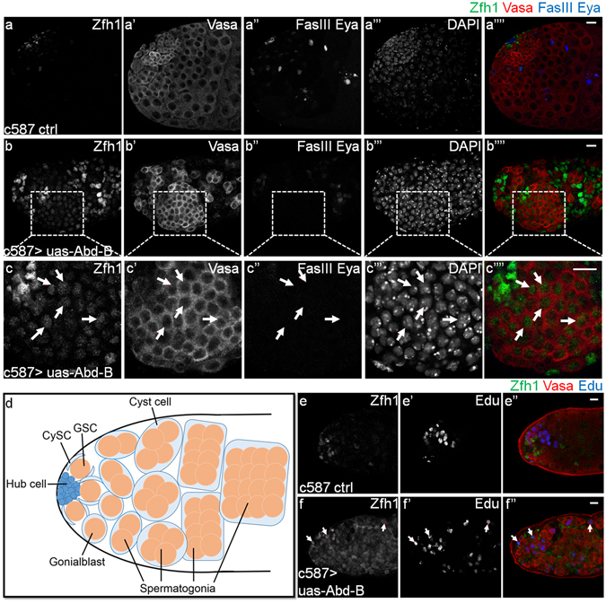
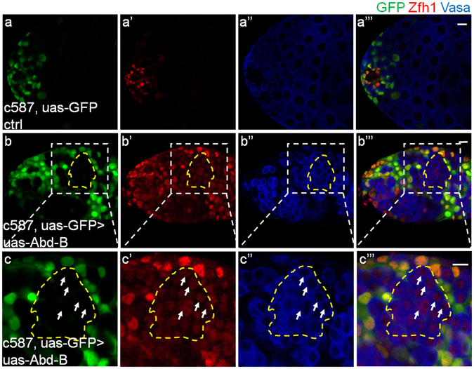
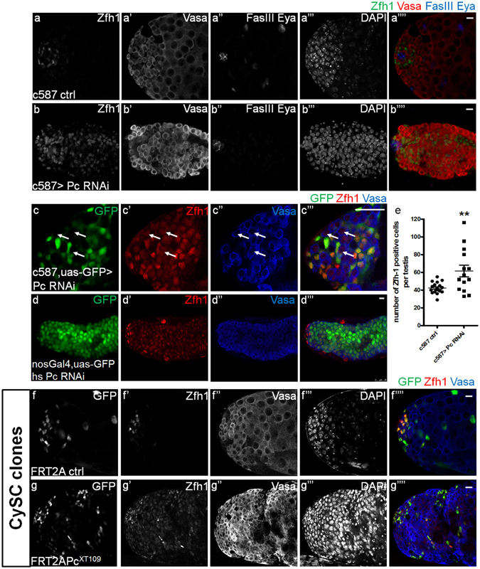
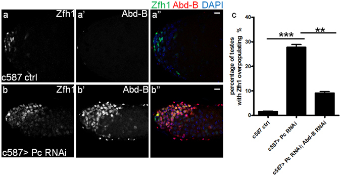
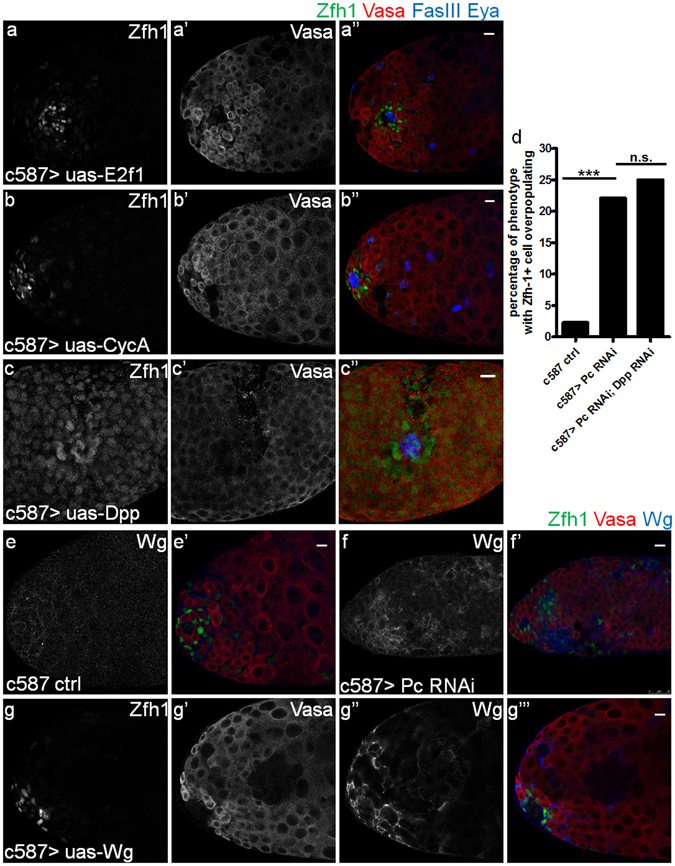
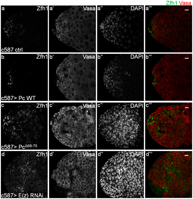
Hox genes play a fundamental role in regulating animal development. However, less is known about their functions on homeostasis maintenance in adult stem cells. Here, we report that the repression of an important axial Hox gene, Abdominal-B (Abd-B), in cyst stem cells (CySCs) is essential for the homeostasis and cell identity maintenance in the adult Drosophila testis. Derepression of Abd-B in CySCs disrupts the proper self-renewal of both germline stem cells (GSCs) and CySCs, and leads to an excessive expansion of early stage somatic cells, which originate from both lineages. We further demonstrate that canonical Polycomb (Pc) and functional pathway of Polycomb group (PcG) proteins are responsible for maintaining the germline cell identity non-autonomously via repressing Abd-B in CySCs in the adult Drosophila testis.


Conflict of interest statement

The authors declare that they have no competing interests.

Figures

Figure 1
Figure 1
Overexpressing Abd-B in CySCs leads to a severe dysfunction in the adult testis. (a–c””) Immunostaining of representative testes showed the CySC and early cyst cell marker Zfh1 (green, locates in the nucleus), germline marker Vasa (red, locates in the cytoplasm), hub cell specific marker FasIII (blue) and differentiated cyst cell specific marker Eya (blue) after Abd-B was overexpressed driven by c587-Gal4 for 15 days at 29 °C. (a-a””) c587 control, (b-b””) c587 > uas-Abd-B, (c-c””) a detailed view of (b-b””). The white arrows indicate cells with both the Zfh1 and Vasa signals. Scale bar, 10 μm. (d) A schematic diagram of the apex of adult Drosophila testis, showing different cell types: post mitotic somatic hub (blue), cyst stem cells (CySCs, light blue), differentiated cyst cells (light blue), germline stem cells (GSCs, brown), gonialblasts (brown) and spermatogonia (brown). A GSC produces a new GSC and a gonialblast through the asymmetric division along with one pair of cyst cells produced through the asymmetric divisions of a pair of CySCs. Then the goniablast continues to differentiate including the transient amplification, growth of spermatocytes, meiosis and the formation of sperm bundles (not shown here). In this process, the cyst cell just elongates its cytoplasm without division. (e-f”) Edu incorporation assay in adult Drosophila testis. The Edu is a nucleoside analog of thymidine and can be incorporated into DNA during active DNA synthesis. This assay was employed to indicate the cell proliferation. Confocal images of representative testes after Abd-B was overexpressed driven by c587-Gal4 for 15 days at 29 °C, showing Zfh1 (green), Vasa (red), Edu (blue). (e-e”) c587 control, (f-f”) c587 > uas-Abd-B. Arrows in (f-f”) indicate the single dividing cells, which locate far away from hub region. Scale bar, 10 μm.
Figure 2
Figure 2
Some Zfh1 positive cells are not derived from cyst cell lineage when Abd-B was overexpressed in CySCs. (a-a”’) Representative testes in the lineage tracing experiments using the c587-Gal4ts, uas-GFP control transgene showing GFP (green), Zfh1 (red) and Vasa (blue) after 20 days’ induction at 29 °C, in which the GFP colocalizes with Zfh1. (b-b”’) c587-Gal4ts, uas-GFP > uas-Abd-B testes showing the GFP (green), Zfh1 (red) and Vasa (blue) after 20 days’ induction at 29 °C, (c-c”’) the detailed view of the white circled part in (b-b”’). Most of the cells in the yellow broken lines are stained with both Zfh1 (red) and Vasa (blue) signals, but not GFP, as the representative cells signed with the white arrows. Scale bar, 10 μm.
Figure 3
Figure 3
Pc promotes cell differentiation and non-autonomously maintains germline cell identity. (a-b””) representative testes showing the Zfh1 (green), Vasa (red), FasIII (blue) and Eya (blue) after RNAi induction for 21 days at 29 °C. (a-a””) c587 control, (b-b””) c587 > Pc RNAi. Scale bar, 10 μm. (c-c”’) Immunostaining of testes for lineage tracing using the c587-Gal4ts, uas-GFP transgene combined with Pc RNAi after induction for 21 days at 29 °C. GFP (green), Zfh1 (red), Vasa (blue). White arrows indicate the GFP negative cells with both Zfh1 and Vasa signals. Scale bar, 10 μm. (d-d”’) Immunostaining of testes using the nosGal4, uas-GFP transgene, which labels the germline cells with GFP, combined with hs Pc RNAi after heat shock for 21 days. GFP (green), Zfh1 (red), Vasa (blue). Scale bar, 10 μm. (e) Quantification of the number of Zfh1 positive cells per testis in c587 ctrl and c587 > Pc RNAi after RNAi induction in 29 °C for 10 days. The total number of testes for each genotype is more than 20. Statistical significance is determined by Student’s t-test. Data are presented as mean ± SEM. **P < 0.01. (f-g””) Immunostaining of testes after clone induction for 15 days. Testes with GFP-positively marked FRT2A clones (f-f””) or FRT2APcXT109 (g-g””) clones are immunostained to show GFP (green), Zfh1 (red), Vasa (Blue). CySC clones (Zfh1+GFP+) are located around the hub region at 15 days ACI in control, CySC clones (Zfh1+GFP+) in FRT2APcXT109 are much more far away from the hub at 15 days ACI. Scale bar, 10 μm.
Figure 4
Figure 4
Pc represses Abd-B to maintain the homeostasis in adult Drosophila testis. (a-b”) Immunostaining of Abd-B expression level in adult Drosophila testes after RNAi induction for 21 days in 29 °C. (a-a”) c587 control, (b-b”) c587 > Pc RNAi. Zfh1(green), Abd-B (red) and DAPI (blue). Scale bar, 10 μm. (c) Quantification of the penetrance of testes with Zfh1 positive cells overpopulating after induction for 21 days at 29 °C. The number of testes scored for each transgene is more than 100. Data are presented as mean ± SEM. Statistical significance is determined by Student’s t-test, ***P < 0.001, **P < 0.01.
Figure 5
Figure 5
E2f1, CycA, Dpp and Wg are not involved in the disorder induced by Pc downregulation. (a-a”) Representative testes with CySC specific E2f1 overexpression in 29 °C for 15 days showing Zfh1 (green), Vasa (red), FasIII (blue) and Eya (blue). (b-b”) Representative testes with CySC specific CycA overexpression in 29 °C for 15 days showing Zfh1 (green), Vasa (red), FasIII (blue) and Eya (blue). (c-c”) Representative testes with CySC specific Dpp overexpression in 29 °C for 10 days showing Zfh1 (green), Vasa (red), FasIII (blue) and Eya (blue). (d) Quantification of percentage of testes with Zfh1 positive cells overpopulating (the number of Zfh1 positive cells is over 60) in the control, c587 > Pc RNAi, c587 > Pc RNAi; Dpp RNAi. The number of flies in each group is more than 50. Statistical significance is determined by Student’s t-test. *** P < 0.001, n.s., not significant. (e-f’) Representative testes showing Zfh1(green), Vasa (red) and Wgless (blue) after RNAi induction for 21 days at 29 °C. Wgless is an important ligand of Wnt signal pathway. (e-e’) c587 control, (f-f’) c587 > Pc RNAi. (g-g”’) Representative testes show Zfh1(green), Vasa (red) and Wgless (blue) in the background of the CySC specific Wglesss overexpression.
Figure 6
Figure 6
The function of Pc is dependent on H3K27me3 in the Drosophila testis. Indicated testes are immunostained to show Zfh1 (green), Vasa (red) after the transgenes were overexpressed driven by CySC specific driver c587-Gal4 for 21 days at 29 °C. (a-a”’) c587 control, (b-b”’) c587 > uas-Pc WT, (c-c”’) c587 > uas-PcΔ69–70, (d-d”’) c587 > E(z) RNAi. Scale bar, 10 μm.

Similar articles

Cited by

References

    1. Spradling A, Fuller MT, Braun RE, Yoshida S. Germline stem cells. Cold Spring Harb Perspect Biol. 2011;3:a002642. doi: 10.1101/cshperspect.a002642. - DOI - PMC - PubMed
    1. Lehmann R. Germline stem cells: origin and destiny. Cell stem cell. 2012;10:729–739. doi: 10.1016/j.stem.2012.05.016. - DOI - PMC - PubMed
    1. Fuller MT, Spradling AC. Male and female Drosophila germline stem cells: two versions of immortality. Science. 2007;316:402–404. doi: 10.1126/science.1140861. - DOI - PubMed
    1. Greenspan LJ, de Cuevas M, Matunis E. Genetics of Gonadal Stem Cell Renewal. Annual review of cell and developmental biology. 2015 - PMC - PubMed
    1. Kawase E, Wong MD, Ding BC, Xie T. Gbb/Bmp signaling is essential for maintaining germline stem cells and for repressing bam transcription in the Drosophila testis. Development. 2004;131:1365–1375. doi: 10.1242/dev.01025. - DOI - PubMed

Publication types

MeSH terms

LinkOut - more resources