. 2017 Jul 11;7(1):5077.
doi: 10.1038/s41598-017-05392-z.

Evaluation of Osteogenesis and Angiogenesis of Icariin in Local Controlled Release and Systemic Delivery for Calvarial Defect in Ovariectomized Rats

Affiliations

Evaluation of Osteogenesis and Angiogenesis of Icariin in Local Controlled Release and Systemic Delivery for Calvarial Defect in Ovariectomized Rats

Yuqiong Wu et al. Sci Rep. .

Abstract

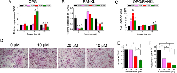
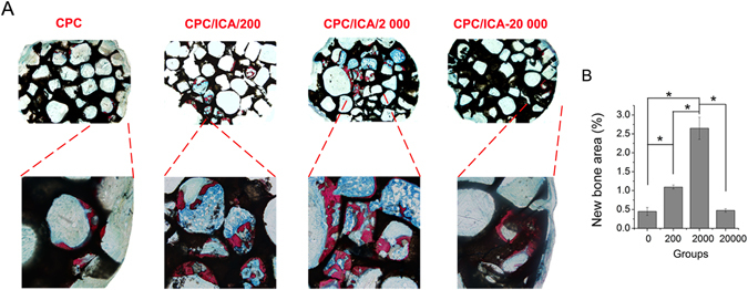
Typically, bone regenerative medicine is applied to repair bone defects in patients with osteoporosis. Meanwhile, there is an urgent need to develop safe and cheap drugs that induce bone formation. Icariin, which is reported to promote the osteogenesis of stem cells in vitro, is the main active component of Herba Epimedii. However, whether icariin could repair bone defects caused by osteoporosis remains unknown. In this study, an osteoporosis model in rats was established by an ovariectomy first, and then, the osteogenic and angiogenic differentiation of bone mesenchymal stem cells (BMSCs) treated with icariin was evaluated. Furthermore, calcium phosphate cement (CPC) scaffolds loaded with icariin were constructed and then implanted into nude mice to determine the optimal construction. To evaluate its osteogenic and angiogenic ability in vivo, this construction was applied to calvarial defect of the ovariectomized (OVX) rats accompanied with an icariin gavage. This demonstrated that icariin could up-regulate the expression of osteogenic and angiogenic genes in BMSCs. Meanwhile, osteoclast formation was inhibited. Moreover, CPC could act as a suitable icariin delivery system for repairing bone defects by enhancing osteogenesis and angiogenesis, while the systemic administration of icariin has an antiosteoporotic effect that promotes bone defect repair.


Conflict of interest statement

The authors declare that they have no competing interests.

Figures

Figure 1
Figure 1
The proliferation, osteoblastic differentiation and angiogenic factor expression effect of icariin on BMSCs. (A) Proliferation of BMSCs treated with icariin at concentrations of 10, 20 and 40 µM. (BF) Real-time PCR analysis of Runx2, Col I, ALP, OPN, OCN, VEGF and ANG1 mRNA in BMSCs treated with icariin at concentrations of 10, 20 and 40 µM. (I) ALP staining of BMSCs after treatment with icariin on days 7 and 10. (J) Quantitative ALP activity of BMSCs treated with icariin measured by the pNPP assay on days 7 and 10. (*p < 0.05, n = 3).
Figure 2
Figure 2
Effect of icariin on the expression of osteoclastic genes and formation of osteoclasts. (A,B) Real-time PCR analysis of OPG and RANKL mRNA in BMSCs treated with icariin at concentrations of 10, 20 and 40 µM. (C) The ratio of OPG/RANKL. (D) Formation of osteoclasts. (E) Number of osteoclasts. (F) Area of osteoclasts. (*p < 0.05, n = 3).
Figure 3
Figure 3
Release of icariin loaded in CPC at concentrations of 200, 2 000 and 20 000 μM, and the morphology, adhesion, and proliferation of BMSCs seeded on CPC with or without icariin. (A) Absolute release amount. (B) Cumulative release of icariin in percent. (C) SEM analysis of BMSCs seeded on samples CPC, CPC/ICA-200, CPC/ICA-2 000, CPC/ICA-20 000 for 2 h and 24 h. Scale bar = 100 μm. (D) The vitality of BMSCs on samples CPC, CPC/ICA-200, CPC/ICA-2 000 and CPC/ICA-20 000.
Figure 4
Figure 4
Osteogenic effect of local icariin on BMSCs in CPC scaffolds implanted in nude mice. On these nondecalcified sections, red areas represent newly formed bone, blue areas belong to collagen fibers, and black areas represent undegraded CPC scaffolds. (A) Histological images of bone formation for group CPC, CPC/ICA-200, CPC/ICA-2 000, and CPC/ICA-20 000 (Line 1: ×40; Line 2: ×100); (B) The percentage of new bone area was assessed by histomorphometric analysis (*p < 0.05).
Figure 5
Figure 5
Sequential fluorescent labeling of TE, AL and CA for CPC, CPC/ICA-2 000, CPC +ICA-ig, and CPC/ICA-2 000 +ICA-ig groups. The images in yellow (TE), red (AL) and green (CA) indicated the rate of bone formation and mineralization at 2, 4 and 6 weeks after the operation, respectively. Merged images of the three fluorochromes for the same group. Scale bar = 100 μm. (B) The percentage of TE, AL and CA staining by histomorphometric analysis (*p < 0.05).
Figure 6
Figure 6
Histological images of newly formed bone in calvarial defects for groups CPC (Group A), CPC/ICA-2 000 (Group B), CPC +ICA-ig (Group C) and CPC/ICA-2 000 +ICA-ig (Group D) that were taken 8 weeks after the operation (Line 1: ×40; Line 2: ×100). On these nondecalcified sections, red areas represent newly formed bone, and black areas belong to undegraded CPC scaffolds. (B) The percentages of new bone area were assessed by histomorphometric analysis (*p < 0.05).
Figure 7
Figure 7
Histological images of newly formed blood vessels in calvarial defects for groups CPC(Group A), CPC/ICA-2 000 (Group B), CPC +ICA-ig (Group C) and CPC/ICA-2 000 +ICA-ig (Group D) that were taken at 8 weeks after the operation. (B,C) The objective surface density and objects number of newly formed blood vessel were assessed by histomorphometric analysis (*p < 0.05).
Figure 8
Figure 8
The induced effect of icariin on the weight (A), serum calcium (B), and serum phosphorus (C) content, and femoral biomechanical parameters (D) in OVX rats (*p < 0.05).
Figure 9
Figure 9
Schematic diagram of depicting the conceptual framework.

Similar articles

Cited by

References

    1. Consensus Development Conference on Osteoporosis. Hong Kong, April 1–2, 1993. Am J Med95, 1S–78S (1993). - PubMed
    1. Khosla S. Increasing options for the treatment of osteoporosis. N Engl J Med. 2009;361:818–20. doi: 10.1056/NEJMe0905480. - DOI - PMC - PubMed
    1. Gorman C, Park A. The truth about hormones. Time. 2002;160:32–9. - PubMed
    1. Li WJ, Tuli R, Huang X, Laquerriere P, Tuan RS. Multilineage differentiation of human mesenchymal stem cells in a three-dimensional nanofibrous scaffold. Biomaterials. 2005;26:5158–66. doi: 10.1016/j.biomaterials.2005.01.002. - DOI - PubMed
    1. Zeng D, et al. Maxillary sinus floor elevation using a tissue-engineered bone with calcium-magnesium phosphate cement and bone marrow stromal cells in rabbits. Tissue Eng Part A. 2012;18:870–81. doi: 10.1089/ten.tea.2011.0379. - DOI - PubMed

Publication types

MeSH terms

LinkOut - more resources