. 2017 Jul 11;7(1):5103.
doi: 10.1038/s41598-017-05394-x.

Dopamine 2 Receptor Activation Entrains Circadian Clocks in Mouse Retinal Pigment Epithelium

Affiliations

Dopamine 2 Receptor Activation Entrains Circadian Clocks in Mouse Retinal Pigment Epithelium

Kenkichi Baba et al. Sci Rep. .

Abstract

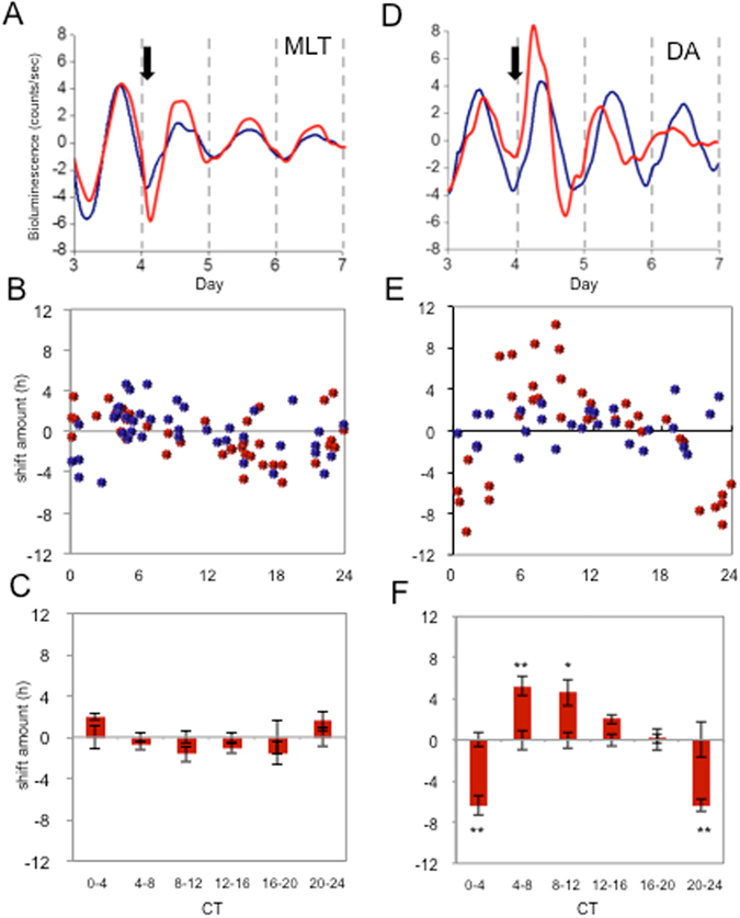
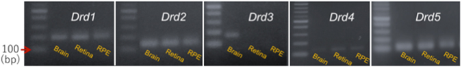
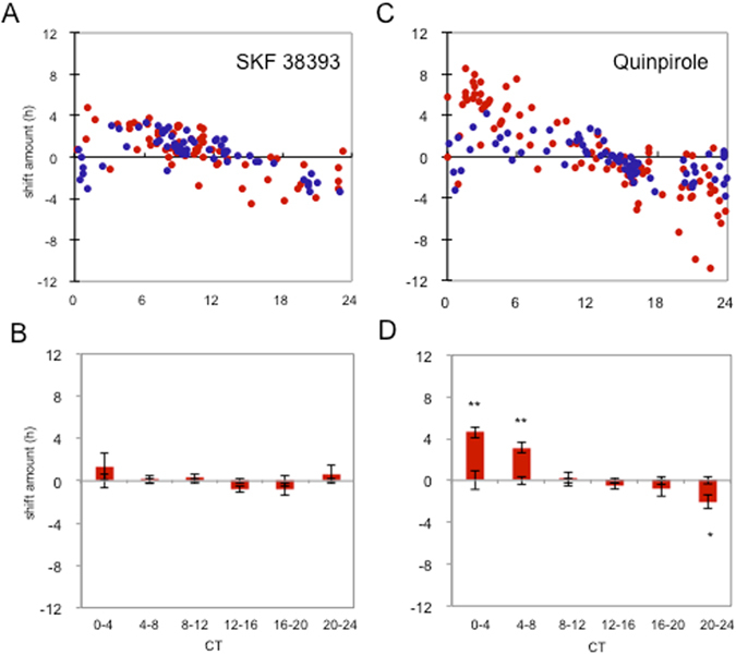
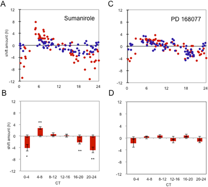
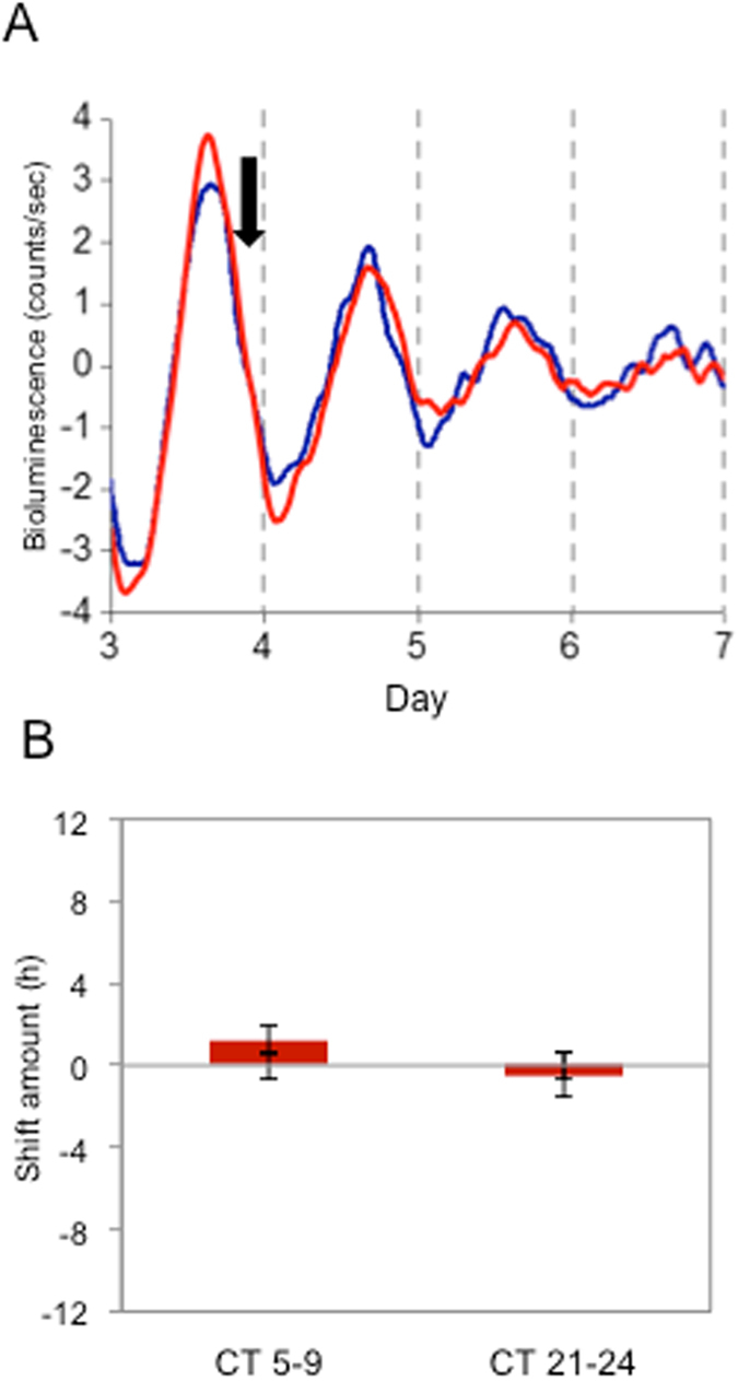
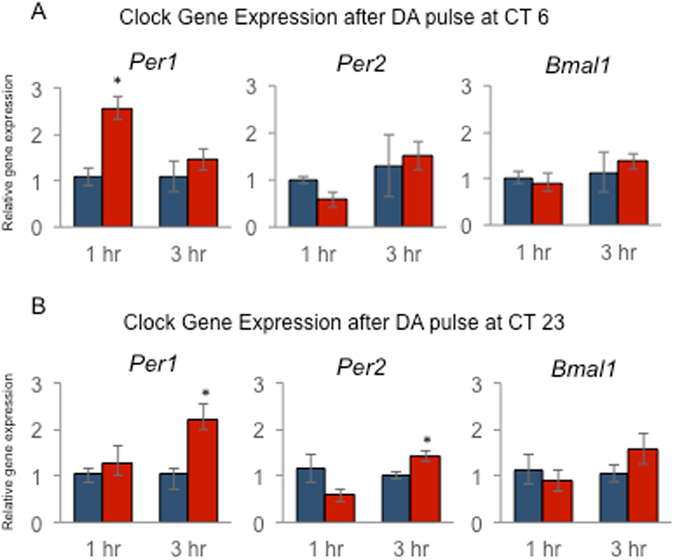
Many of the physiological, cellular, and molecular rhythms that are present within the eye are under the control of circadian clocks. Experimental evidence suggests that the retinal circadian clock, or its output signals (e.g., dopamine and melatonin), may contribute to eye disease and pathology. We recently developed a retinal pigment ephithelium (RPE)-choroid preparation to monitor the circadian clock using PERIOD2 (PER2)::LUC knock-in mouse. In this study we report that dopamine, but not melatonin, is responsible for entrainment of the PER2::LUC bioluminescence rhythm in mouse RPE-choroid. Dopamine induced phase-advances of the PER2::LUC bioluminescence rhythm during the subjective day and phase-delays in the late subjective night. We found that dopamine acts exclusively through Dopamine 2 Receptors to entrain the circadian rhythm in PER2::LUC bioluminescence. Finallly, we found that DA-induced expression of core circadian clock genes Period1 and Period2 accompanied both phase advances and phase delays of the RPE-choroid clock, thus suggesting that - as in other tissues - the rapid induction of these circadian clock genes drives the resetting process. Since the RPE cells persist for the entire lifespan of an organism, we believe that RPE-choroid preparation may represent a new and unique tool to study the effects of circadian disruption during aging.


Conflict of interest statement

The authors declare that they have no competing interests.

Figures

Figure 1
Figure 1
Dopamine phase-shifts PER2::LUC bioluminescence rhythms in mouse RPE-choroid. DA or MLT were added to the culturing media after the third peak of the RPE-choroid PER2::LUC bioluminescence rhythm. The representative data shows application of MLT at CT 14 did not phase-shift the RPE-choroid bioluminescence PER2::LUC rhythm (A). On the other hand, DA application at CT 8 phase-shifted the RPE-choroid bioluminescence rhythm (D). The blue traces indicate controls (vehicle treated) while red traces indicate MLT (A) or DA (D) treated RPE-choroid cultures. The black arrows indicate time of the drug or vehicle (Veh) treatments (A and D). The amount of phase-shift for each individual RPE-choroid rhythm was plotted to create a PRC (B and E). Blue circles indicate cultures treated with Veh and red circles indicate culture treated with either MLT (B) or DA (E). Data were divided into 6 bins at 4-hour intervals for statistical analysis. Data were then used to calculate the phase change of MLT or DA versus their vehicle controls. Bars show the mean amount of phase change from controls and error bars show ±SEM for experimental groups. Error bars from x axis show ±SEM for control groups. MLT did not phase-shift the RPE-choroid PER2::LUC bioluminescence rhythm (n = 4–14 for each bin, Two way ANOVA, p > 0.1, (C) whereas DA significantly phase-shifted PER2::LUC rhythm (Two way ANOVA following Tukey tests, *p < 0.05, **p < 0.01, n = 6–8 for each bin, (F).
Figure 2
Figure 2
Expression of dopamine receptors in the brain, retina, and RPE. Agarose gel electrophoresis of PCR amplicons specific to D1R, D2R, D3R, D4R or D5R transcripts in brain, retina and RPE. D3R mRNA was present in the brain (positive control), but was not amplified in the retina (negative control) or RPE. The electrophoresis bands matched the expected amplicon size.
Figure 3
Figure 3
Effects of D1-like agonist (SKF38393) and D2-like agonist (Quinpirole) on PER2::LUC bioluminescence rhythm. 50 µM of SKF38393 did not phase-shift the PER2::LUC bioluminescence rhythm (A and B), whereas quinpirole significantly phase-advanced PER2::LUC bioluminescence rhythm at CT 0–4 and CT 4–8 hrs and phase-delayed when applied at CT 20–24 (C and D). Blue circles indicate cultures treated with vehicle and red circles indicate culture treated with active compounds. Data were divided to 6 bins at 4-hour intervals for statistical analysis (Two-way ANOVA following Tukey tests, *p < 0.05). n = 3–21 for each bin (B and D). Data were then used to calculate the phase change of drug treated versus their vehicle controls. Bars show the mean amount of phase change from controls and error bars show ±SEM for experimental groups. Error bars from x axis show ±SEM for control groups.
Figure 4
Figure 4
Effects of D2R (Sumanirole) and D4R (PD168077) agonists on the PER2::LUC bioluminescence rhythm. Sumanirole of 1 µM significantly phase-delayed PER2::LUC bioluminescence rhythm at CT 0–4, CT 16–20, and CT 20–24 h. Sumanirole significantly phase-advanced the bioluminescence rhythm at CT 4–8 (A and B). PD166077 did not phase-shift the PER2::LUC bioluminescence rhythms (C and D). Blue circles indicate cultures treated with vehicle and red circles indicate culture treated with active compounds (A and C). Data were divided to 6 bins at 4-hour intervals for statistical analysis (Two-way ANOVA following Tukey tests, *p < 0.05) n = 6–21 for each bin (B and D). Data were then used to calculate the phase change of drug treated versus their vehicle controls. Bars show the mean amount of phase change from controls and error bars show ±SEM for experimental groups. Error bars from x axis show ±SEM for control groups.
Figure 5
Figure 5
DA did not induce phase-shifts in the RPE-choroid of PER2::LUC bioluminescence rhythms in D2R−/−PER2::LUC mice. Representative example shows that DA treatment at CT 24 did not phase-shift D2R−/−PER2::LUC bioluminescence rhythm (A). The blue trace indicates a control (Veh treated) and red trace indicates a DA treated RPE-choroid culture. The black arrow indicates time of the drug or vehicle treatment (A). DA treated RPE-choroid cultures at CT 5–9 and at CT 21–24 were averaged for statistical analysis. Data were used to calculate the phase change of D2R−/− versus controls. Bars show the mean amount of phase change from controls and error bars show ±SEM for experimental groups. Error bars from x axis show ±SEM for control groups. No phase-shifts were observed in the D2R−/−PER2::LUC RPE-choroid rhythms at either time points (B) t-test, n = 6 for DA and Veh for each time point).
Figure 6
Figure 6
DA treatment increases Per1 and Per2 mRNA in RPE-choroid. RPE-choroid cultures were prepared and treated with DA or vehicle at ZT6 (A, advance) or ZT 23 (B, delay) as indicated above, followed by collection for Q-PCR analysis of the indicated mRNAs at 1 or 3 hour intervals. Expression data were normalized using 18S, and are plotted relative to vehicle controls. Blue bars indicate mean ± SEM of vehicle control. Red bars indicate mean ± SEM of DA treated. *Indicates p < 0.05 (t-test) compared to vehicle controls (n = 3–6 in each group).

References

    1. Tosini G, Menaker M. Circadian rhythms in cultured mammalian retina. Science. 1996;272:419–421. doi: 10.1126/science.272.5260.419. - DOI - PubMed
    1. Tosini G, Menaker M. The clock in the mouse retina: melatonin synthesis and photoreceptor degeneration. Brain Res. 1998;789:221–228. doi: 10.1016/S0006-8993(97)01446-7. - DOI - PubMed
    1. McMahon DG, Iuvone PM, Tosini G. Circadian organization of the mammalian retina: from gene regulation to physiology and diseases. Prog Retin Eye Res. 2014;39:58–76. doi: 10.1016/j.preteyeres.2013.12.001. - DOI - PMC - PubMed
    1. Besharse JC, McMahon DG. The Retina and Other Light-sensitive Ocular Clocks. J. Biol. Rhythms. 2016;31:223–243. doi: 10.1177/0748730416642657. - DOI - PMC - PubMed
    1. Yoo SH, et al. PERIOD2::LUCIFERASE real-time reporting of circadian dynamics reveals persistent circadian oscillations in mouse peripheral tissues. Proc. Natl. Acad. Sci. USA. 2004;101:5339–5346. doi: 10.1073/pnas.0308709101. - DOI - PMC - PubMed

Publication types

LinkOut - more resources