Review
. 2017 Sep;18(9):533-547.
doi: 10.1038/nrm.2017.60. Epub 2017 Jul 12.

Genes and molecular pathways underpinning ciliopathies

Affiliations
Review

Genes and molecular pathways underpinning ciliopathies

Jeremy F Reiter et al. Nat Rev Mol Cell Biol. 2017 Sep.

Abstract

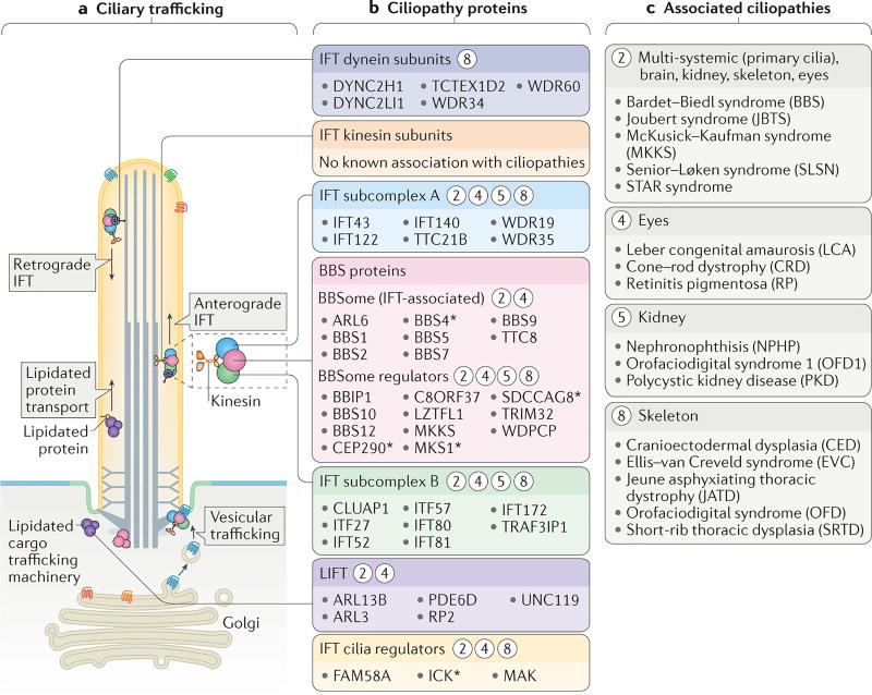
Motile and non-motile (primary) cilia are nearly ubiquitous cellular organelles. The dysfunction of cilia causes diseases known as ciliopathies. The number of reported ciliopathies (currently 35) is increasing, as is the number of established (187) and candidate (241) ciliopathy-associated genes. The characterization of ciliopathy-associated proteins and phenotypes has improved our knowledge of ciliary functions. In particular, investigating ciliopathies has helped us to understand the molecular mechanisms by which the cilium-associated basal body functions in early ciliogenesis, as well as how the transition zone functions in ciliary gating, and how intraflagellar transport enables cargo trafficking and signalling. Both basic biological and clinical studies are uncovering novel ciliopathies and the ciliary proteins involved. The assignment of these proteins to different ciliary structures, processes and ciliopathy subclasses (first order and second order) provides insights into how this versatile organelle is built, compartmentalized and functions in diverse ways that are essential for human health.


Figures

Figure 1
Figure 1. Structures and functions of motile and non-motile cilia
All cilia extend from a basal body that typically consists of triplet microtubules, and subdistal and distal appendages. Distal appendages (also known as transition fibres) tether the basal body to the base of the ciliary membrane. Immediately distal to the basal body is the transition zone, which contains doublet microtubules that are connected to the ciliary membrane via Y-shaped structures. Axonemes (the ciliary backbone) are composed of doublet microtubules. In motile cilia, axonemes usually contain associated structures and proteins (for example, the central pair and axonemal dyneins) that are required for ciliary motility. Nodal cilia are an exception as they are motile but lack a central pair of microtubules. Cilia may contain additional subdomains, including singlet microtubules at the distal end, and regions with specific protein compositions or functions (for example, the inversin domain (INV; involved in signalling). Key cell signalling functions and roles in motility are summarized. PKD, polycystin.
Figure 2
Figure 2. Dysfunctions in motile and/or non-motile cilia cause ciliopathies that encompass most human organ systems
The figure shows the different organ systems or tissues that are affected in diverse ciliopathies, and the principle phenotypic manifestations of the disease in each organ. Ciliopathies that are caused primarily by defects in motile cilia are shown in orange, those that result from defects in non-motile (primary) cilia are shown in blue and those associated with defects in both types of cilia are shown in green. NPHP, nephronophthisis; PKD, polycystic kidney disease.
Figure 3
Figure 3. Structural and functional features of motile and sensory cilia are associated with ciliopathies
a | The major structures of motile and non-motile cilia (also see FIG. 1). b | Major sites of action for ciliopathy-associated proteins that are components of motile cilia (motility apparatus or transcription factors required for the generation of motile cilia) and sensory cilia (axonemal and signalling proteins, ciliary tip proteins or inversin (INV) compartment proteins). The asterisks indicate proteins that are also localized to other ciliary regions during ciliogenesis (shown in FIG. 4) or ciliary trafficking (shown in FIG. 5). Circled numbers indicate one or more ciliopathies that result from defects in the different ciliary compartments and proteins. c | Ciliopathies grouped into major categories that are associated with the proteins and ciliary regions shown in part b.
Figure 4
Figure 4. Ciliogenesis and ciliary compartmentalization are associated with ciliopathies
a | The early steps of ciliogenesis. A mother centriole matures into a basal body and migrates towards the plasma membrane. The basal body distal appendages interact either directly with the plasma membrane, or via an intermediary ciliary vesicle (as shown), and the basal body-associated membrane becomes the incipient ciliary membrane. The transition zone is the first ultrastructure of the cilium to form. Centriolar satellites have a role in ciliogenesis, potentially as an intermediate storage compartment for ciliogenic proteins. b | Ciliopathy proteins associated with different sub-compartments of the basal body, the centriolar satellites or the ciliary apparatus during and/or after ciliogenesis. Circled numbers indicate which ciliopathies (listed in part c) result from defects in these sub-compartments, as well as the organs, tissues or physiological functions that are affected. The asterisks indicate proteins that are also localized to other ciliary regions during ciliogenesis or ciliary trafficking (shown in FIG. 5). c | Ciliopathies grouped into major categories that are associated with the proteins and ciliary compartments shown in part b.
Figure 5
Figure 5. Links between ciliary trafficking and ciliopathies
a | The functional components of two ciliary trafficking pathways: intraflagellar transport (IFT) and lipidated protein intraflagellar targeting (LIFT). Ciliary proteins are trafficked from the Golgi or cytosol to the base of the cilium, after which they are transported into the ciliary compartment. IFT modules that mediate trafficking include anterograde (kinesin-2) and retrograde (dynein-2) motors, IFT subcomplexes A and B, and an accessory module that contains Bardet–Biedl syndrome (BBS) proteins (the BBSome). b | Ciliopathy proteins that constitute, or are regulators of, the IFT and LIFT trafficking systems. Circled numbers indicate which ciliopathies (listed in part c) result from defects in these ciliary trafficking components. The asterisks indicate proteins that are also localized to other ciliary regions during ciliogenesis (shown in FIG. 4) or ciliary trafficking. c | Ciliopathies that result from defects in ciliary trafficking grouped into categories according to the tissues affected.

References

    1. Carvalho-Santos Z, Azimzadeh J, Pereira-Leal JB, Bettencourt-Dias M. Evolution: Tracing the origins of centrioles, cilia, and flagella. J Cell Biol. 2011;194:165–175. - PMC - PubMed
    1. Fisch C, Dupuis-Williams P. Ultrastructure of cilia and flagella back to the future! Biol Cell. 2011;103:249–270. - PubMed
    1. Nachury MV. How do cilia organize signalling cascades. Philos Trans R Soc Lond B Biol Sci. 2014;369 - PMC - PubMed
    1. Reiter JF, Blacque OE, Leroux MR. The base of the cilium: roles for transition fibres and the transition zone in ciliary formation, maintenance and compartmentalization. EMBO Rep. 2012;13:608–618. - PMC - PubMed
    1. Garcia-Gonzalo FR, Reiter JF. Scoring a backstage pass: mechanisms of ciliogenesis and ciliary access. J Cell Biol. 2012;197:697–709. - PMC - PubMed

Substances