. 2017 Jul 11;7(1):5121.
doi: 10.1038/s41598-017-04974-1.

Naringenin Impairs Two-Pore Channel 2 Activity And Inhibits VEGF-Induced Angiogenesis

Affiliations

Naringenin Impairs Two-Pore Channel 2 Activity And Inhibits VEGF-Induced Angiogenesis

Irene Pafumi et al. Sci Rep. .

Abstract

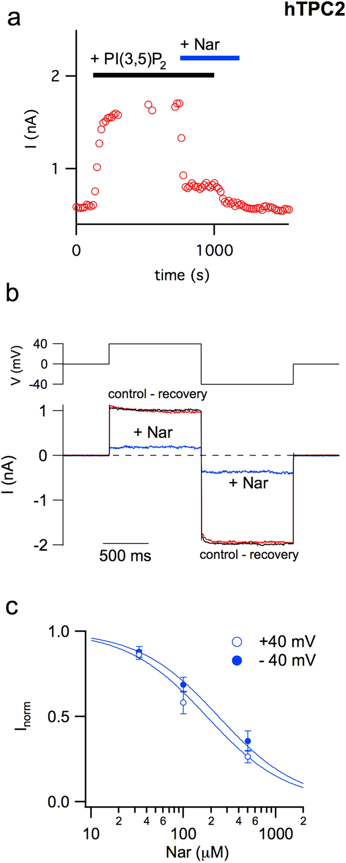
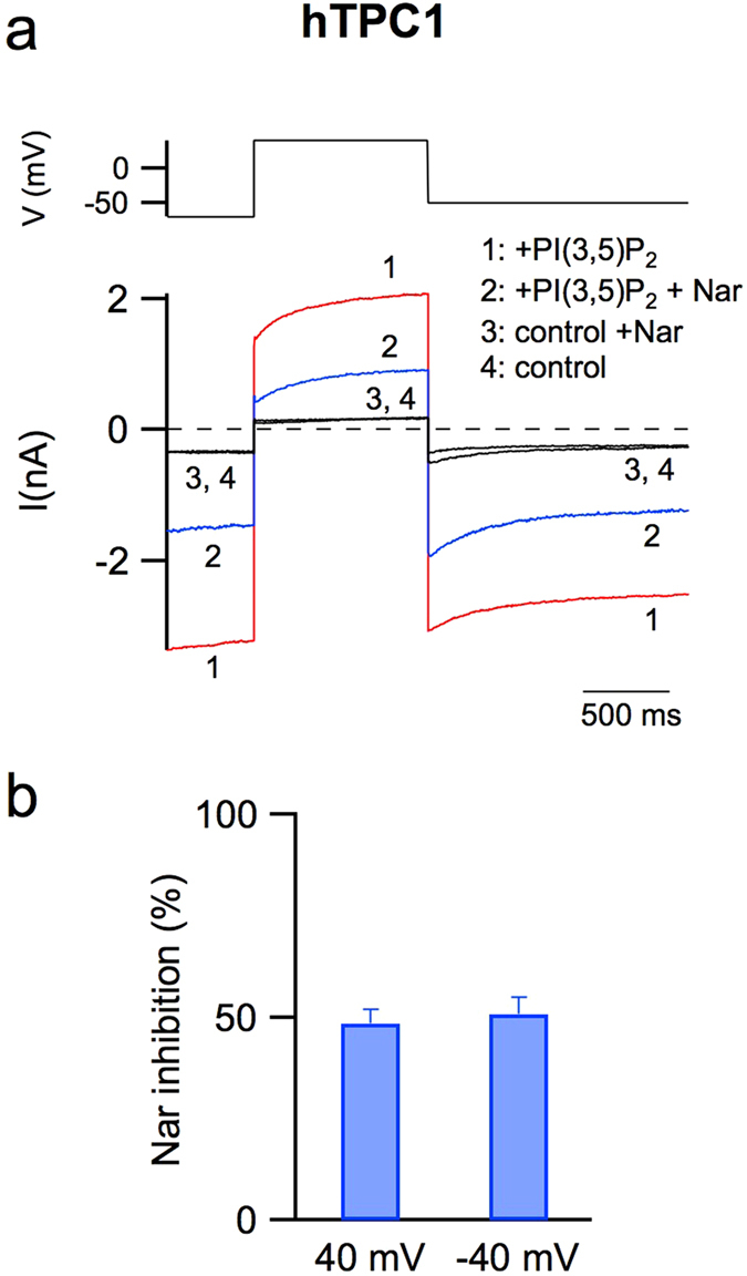
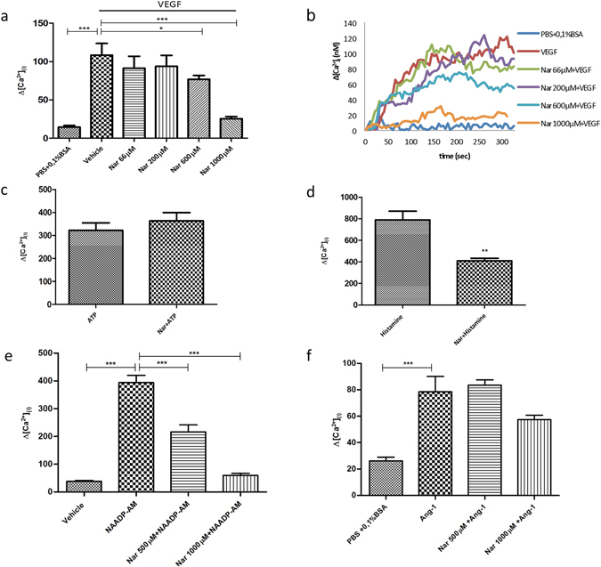
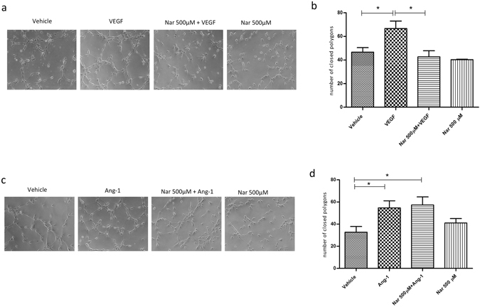
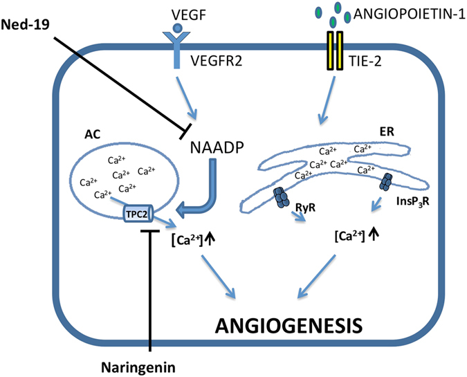
Our research introduces the natural flavonoid naringenin as a novel inhibitor of an emerging class of intracellular channels, Two-Pore Channel 2 (TPC2), as shown by electrophysiological evidence in a heterologous system, i.e. Arabidopsis vacuoles lacking endogenous TPCs. In view of the control exerted by TPC2 on intracellular calcium signaling, we demonstrated that naringenin dampens intracellular calcium responses of human endothelial cells stimulated with VEGF, histamine or NAADP-AM, but not with ATP or Angiopoietin-1 (negative controls). The ability of naringenin to impair TPC2-dependent biological activities was further explored in an established in vivo model, in which VEGF-containing matrigel plugs implanted in mice failed to be vascularized in the presence of naringenin. Overall, the present data suggest that naringenin inhibition of TPC2 activity and the observed inhibition of angiogenic response to VEGF are linked by impaired intracellular calcium signaling. TPC2 inhibition is emerging as a key therapeutic step in a range of important pathological conditions including the progression and metastatic potential of melanoma, Parkinson's disease, and Ebola virus infection. The identification of naringenin as an inhibitor of TPC2-mediated signaling provides a novel and potentially relevant tool for the advancement of this field of research.


Conflict of interest statement

The authors declare that they have no competing interests.

Figures

Figure 1
Figure 1
Naringenin inhibits the activity of the human TPC2 channel. (a) Time course of current amplitude recorded in response to bath application of 330 nM PI(3,5)P2 (upper black bar) before and after adding 500 μM Nar (upper blue bar). Each point represents the steady-state current at +40 mV. (b) Currents mediated by hTPC2 and induced by the application of 330 nM PI(3,5)P2 in the cytosolic bath solution were recorded at +40 and −40 mV (shown in the upper voltage profile) in the absence or presence of 500 μM cytosolic Nar. For the sake of clarity, background currents in the absence of the phosphoinositide were directly subtracted. (c) Dose–response analysis of Nar inhibition. Inorm, the normalised current, was obtained by the ratio of currents recorded respectively in the presence and absence of cytosolic Nar. Both at +40 and −40 mV, normalised currents at different Nar concentrations were fitted with a Michaelis–Menten function (continuous lines). Data from 3 (Nar 33 μM), 6 (Nar 100 μM) and 9 (Nar 500 μM) different vacuoles were shown as mean ± s.e.m. Values at positive and negative voltages, at a defined Nar concentration, were not significant (P > 0.2); values at different Nar concentrations and at a defined voltage (+40 or −40 mV), were statistically significant (at least P < 0.01).
Figure 2
Figure 2
The activity of the human TPC1 channel is inhibited by naringenin. (a) Currents (lower panel) recorded in control conditions (trace 4) or adding in the cytosolic bath solution: 90 nM PI(3,5)P2 (trace 1), 90 nM PI(3,5)P2 and 500 μM Nar (trace 2), 500 μM Nar (trace 3). In the upper panel the voltage profile is shown. (b) Percentage of current inhibition induced by 500 μM Nar added in the cytosolic solution at −40 and + 40 mV. Data from 6 different vacuoles, shown as mean ± s.e.m., were not statistically significant (P > 0.6).
Figure 3
Figure 3
Naringenin inhibits Ca2+ release from HUVEC acidic stores under VEGF or NAADP-AM stimulation. Ca2+ imaging experiments. (a,b) Cells were pretreated for 30 min with different concentrations of Nar and then stimulated with 100 ng/ml VEGF; (a) Bar chart showing maximum Ca2+ concentrations; (b) Changes in Ca2+ levels shown as representative traces. (c,d) Cells were pretreated with 1000 µM Nar for 30 min, then stimulated with (c) 10 µM ATP (negative control) or (d) 100 µM histamine (positive control). (e,f) Cells were pretreated with 500 µM or 1000 µM Nar and then stimulated with (e) 500 nM NAADP-AM or (f) 100 ng/ml Ang-1. Data in bar charts were from 3 independent experiments, n = 41–180 cells. *P < 0.05; **P < 0.01; ***P < 0.001. Neither 500 µM Nar nor 1000 µM Nar significantly inhibited Ang-1 mediated Ca2+ release (P > 0.2).
Figure 4
Figure 4
Naringenin impairs VEGF-induced vessel formation in vitro. (a) Representative images of one of three independent experiments. HUVECs were plated in Matrigel-coated dishes and incubated for 3–4 h in EBM-2 + 2% FBS supplemented or not with VEGF or Nar, or in medium containing both VEGF and Nar. (c) Representative images of one of three independent experiments. HUVECs were plated in Matrigel-coated dishes and incubated for 2–3 h in EGM-2 supplemented or not with Ang-1 or Nar, or in medium containing both Ang-1 and Nar. (b,d) Quantitative evaluation of tube formation as the number of closed polygons formed in 6 fields for each experimental condition for VEGF (b) and Ang-1 (d). Data in bar charts represent mean ± s.e.m. of three independent experiments. *P < 0.05.
Figure 5
Figure 5
Simplified representation of differential Ca2+ signalling pathways involved in the control of angiogenesis by VEGF and Ang-1. Calcium signalling is inhibited by Nar when mediated by TPC2 but not by RyR/InsP3R. AC: acidic compartments, ER: endoplasmic reticulum.
Figure 6
Figure 6
Naringenin impairs in vivo vascularization induced by VEGF in C57BL/6 mice. In vivo vessel formation was assessed after subcutaneous injection of 5 weeks old male/female C57BL/6 mice with Matrigel plugs containing either vehicle or VEGF or VEGF plus 1000 μM Nar. Five days after injection the mice were sacrificed and plug vascularization was evaluated both macroscopically, as shown in two representative images (a) and as hemoglobin content expressed as absorbance (OD)/1 g matrigel plug (b); values from three independent experiments are expressed as mean ± s.e.m. *P < 0.05. n = 14–15 plugs for each experimental condition.

References

    1. Giovannucci E. Tomatoes, tomato-based products, lycopene, and cancer: review of the epidemiologic literature. J. Natl. Cancer Inst. 1999;91:317–331. doi: 10.1093/jnci/91.4.317. - DOI - PubMed
    1. Alam MA, et al. Effect of citrus flavonoids, naringin and naringenin, on metabolic syndrome and their mechanisms of action. Adv. Nutr. Bethesda Md. 2014;5:404–417. doi: 10.3945/an.113.005603. - DOI - PMC - PubMed
    1. Mir IA, Tiku AB. Chemopreventive and therapeutic potential of ‘naringenin,’ a flavanone present in citrus fruits. Nutr. Cancer. 2015;67:27–42. doi: 10.1080/01635581.2015.976320. - DOI - PubMed
    1. Li Q, et al. Naringenin exerts anti-angiogenic effects in human endothelial cells: Involvement of ERRα/VEGF/KDR signaling pathway. Fitoterapia. 2016;111:78–86. doi: 10.1016/j.fitote.2016.04.015. - DOI - PubMed
    1. Lee SH, et al. Cholesterol-lowering activity of naringenin via inhibition of 3-hydroxy-3-methylglutaryl coenzyme A reductase and acyl coenzyme A:cholesterol acyltransferase in rats. Ann. Nutr. Metab. 1999;43:173–180. doi: 10.1159/000012783. - DOI - PubMed

Publication types

MeSH terms

LinkOut - more resources