. 2017 Jul 11;7(1):5157.
doi: 10.1038/s41598-017-05358-1.

Distinct Properties of Long-Term Potentiation in the Dentate Gyrus along the Dorsoventral Axis: Influence of Age and Inhibition

Affiliations

Distinct Properties of Long-Term Potentiation in the Dentate Gyrus along the Dorsoventral Axis: Influence of Age and Inhibition

An Schreurs et al. Sci Rep. .

Abstract

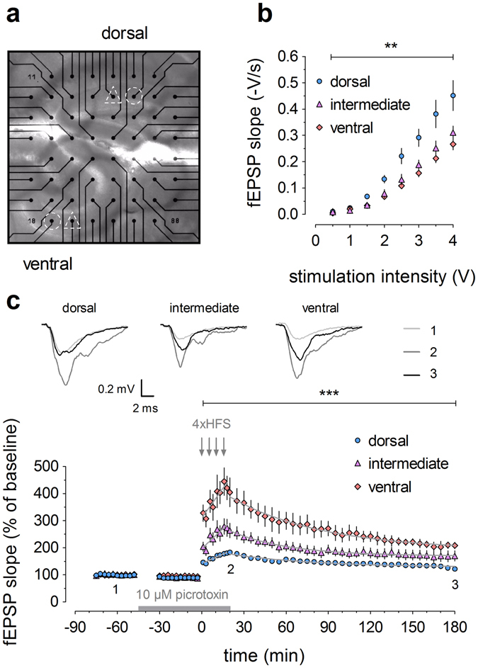
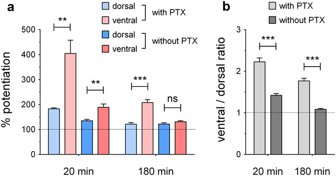
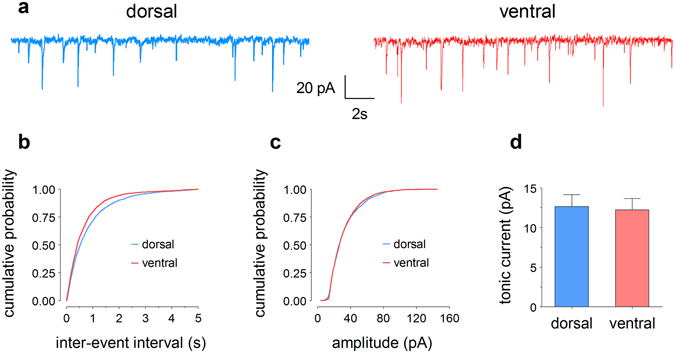
The hippocampus is important for spatial navigation, episodic memory and affective behaviour. Increasing evidence suggests that these multiple functions are accomplished by different segments along the dorsal-ventral (septal-temporal) axis. Long-term potentiation (LTP), the best-investigated cellular correlate of learning and memory, has distinct properties along this axis in the CA1 region, but so far, little is known about longitudinal differences in dentate gyrus (DG). Therefore, here we examined potential dorsoventral differences in DG-LTP using in vitro multi-electrode array recordings. In young mice, we found higher basal synaptic transmission in the dorsal DG, while the LTP magnitude markedly increased towards the ventral pole. Strikingly, these differences were greatly reduced in slices from middle-aged mice. Short-term plasticity, evaluated by paired-pulse ratios, was similar across groups. Recordings in the presence and absence of GABAA-receptor blocker picrotoxin suggested a higher inhibitory tone in the ventral DG of young mice, confirmed by an increased frequency of miniature inhibitory postsynaptic currents. Our findings support the view that the hippocampus contains discrete functional domains along its dorsoventral axis and demonstrate that these are subject to age-dependent changes. Since these characteristics are presumably conserved in the human hippocampus, our findings have important clinical implications for hippocampus- and age-related disorders.


Conflict of interest statement

The authors declare that they have no competing interests.

Figures

Figure 1
Figure 1
Young mice (2–3 months old) exhibit distinct electrophysiological properties in dorsal, intermediate and ventral dentate gyrus. (a) Image of a dorsal (top) and ventral (bottom) hippocampal slice positioned simultaneously on the multi-electrode array (MEA), zoomed in on the dentate gyrus. Selected stimulation electrodes (triangles) target the medial perforant path. Recording channels of interest (circles) are also indicated. (b) Input/output relationships of dorsal (n = 9), intermediate (n = 6) and ventral (n = 6) hippocampal slices indicate that basal synaptic transmission gradually decreases along the dorsoventral axis. (c) High-frequency stimulation (HFS; four trains of 1 s duration at 100 Hz) in dorsal (n = 9), intermediate (n = 8) and ventral (n = 6) slices induces LTP with a higher magnitude and distinct induction and decay kinetics in the ventral compared to intermediate and dorsal DG. Inset shows representative traces of field excitatory postsynaptic potential (fEPSP) for baseline, 20 min post-HFS and 180 min post-HFS, as indicated by numbers 1–3. 10 μM picrotoxin was added to the ACSF after baseline and until the end of HFS, as indicated by the bar. The grey lines superimposed on the data points denote the best-fitted functions for the induction of potentiation and subsequent decay. See Supplementary Table S1 for all equation parameters. Two-way RM-ANOVA was used for statistical analysis (** indicates p < 0.01 and *** indicates p < 0.001).
Figure 2
Figure 2
Distinct electrophysiological properties in dorsal and ventral dentate gyrus of young (a–c) and middle-aged (d–f) mice. (a,d) Input/output curves show a dorsoventral gradient in young (sample sizes: dorsal = 11, ventral = 10) but not in middle-aged mice (dorsal = 9, ventral = 7). (b,e) Paired-pulse ratios are similar between dorsal and ventral DG, both in young (dorsal = 11, ventral = 9) and middle-aged mice (dorsal = 9, ventral = 7). (c) Long-term potentiation in young mice (dorsal = 7, ventral = 9) is characterized by dorsoventral variation in both the induction and maintenance phase. (f) In middle-aged mice (dorsal = 8, ventral = 6), only the induction phase shows dorsoventral differences, whereafter the curves of dorsal and ventral LTP rapidly converge. Representative signal traces are shown for baseline, 20 min post-HFS and 180 min post-HFS, as indicated by numbers 1–3. In this series of recordings, no picrotoxin was used. The grey lines superimposed on the data points represent the best-fitted functions for the induction and decay phases. See Supplementary Tables S2 and S3 for all equation parameters. RM-ANOVA was used for statistical analysis (* indicates p < 0.05 and ** indicates p < 0.01).
Figure 3
Figure 3
Picrotoxin (PTX) has a stronger potentiating effect on the ventral dentate gyrus in young mice (2–3 months old). (a) Levels of potentiation obtained at 20 and 180 min after the start of HFS, for dorsal and ventral slices, with or without the addition of PTX to the recording solution during induction (as can be derived from Figs 1c and 2c, respectively). (b) Comparison of mean ventral/dorsal ratios, calculated based on all possible ratios, for both conditions at 20 and 180 min. Unpaired t tests with Welch’s correction were used for statistical comparisons (** indicates p < 0.01 and *** indicates p < 0.001).
Figure 4
Figure 4
Whole-cell patch-clamp recordings reveal differences in inhibitory properties between dorsal and ventral DG granule cells. (a) Representative traces of miniature inhibitory postsynaptic currents (mIPSCs) of granule cells in slices from dorsal (blue) and ventral (red) hippocampus. (b) Cumulative probability curves indicate significantly shorter median inter-event intervals, i.e. a higher frequency of mIPSCs, in the ventral (409 ms) compared to dorsal DG (524 ms). (c) In contrast, the median mIPSC amplitudes are not different (dorsal: 26.6 pA, ventral: 26.9 pA). (d) Tonic currents in dorsal and ventral granule cells, calculated as the difference between the holding current in the absence and presence of 50 mM bicuculline, are almost identical (dorsal: 12.6 ± 1.5 pA, ventral: 12.2 ± 1.4 pA).

References

    1. Fanselow MS, Dong H-W. Are the Dorsal and Ventral Hippocampus Functionally Distinct Structures? Neuron. 2010;65:7–19. doi: 10.1016/j.neuron.2009.11.031. - DOI - PMC - PubMed
    1. Strange BA, Witter MP, Lein ES, Moser EI. Functional organization of the hippocampal longitudinal axis. Nat. Rev. Neurosci. 2014;15:655–669. doi: 10.1038/nrn3785. - DOI - PubMed
    1. Moser MB, Moser EI, Forrest E, Andersen P, Morris RG. Spatial learning with a minislab in the dorsal hippocampus. Proc. Natl. Acad. Sci. 1995;92:9697–9701. doi: 10.1073/pnas.92.21.9697. - DOI - PMC - PubMed
    1. Tovote P, Fadok JP, Lüthi A. Neuronal circuits for fear and anxiety. Nat. Rev. Neurosci. 2015;16:317–331. doi: 10.1038/nrn3945. - DOI - PubMed
    1. Kheirbek MA, et al. Differential Control of Learning and Anxiety along the Dorsoventral Axis of the Dentate Gyrus. Neuron. 2013;77:955–968. doi: 10.1016/j.neuron.2012.12.038. - DOI - PMC - PubMed

Publication types