. 2017 Jul 12;6(7):e62.
doi: 10.1038/emi.2017.49.

Modelling person-to-person transmission in an Enterovirus A71 orally infected hamster model of hand-foot-and-mouth disease and encephalomyelitis

Affiliations

Modelling person-to-person transmission in an Enterovirus A71 orally infected hamster model of hand-foot-and-mouth disease and encephalomyelitis

Win Kyaw Phyu et al. Emerg Microbes Infect. .

Abstract

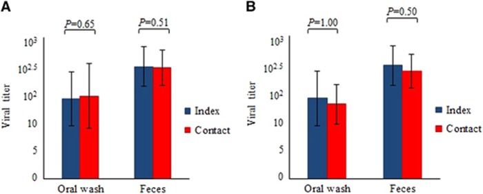
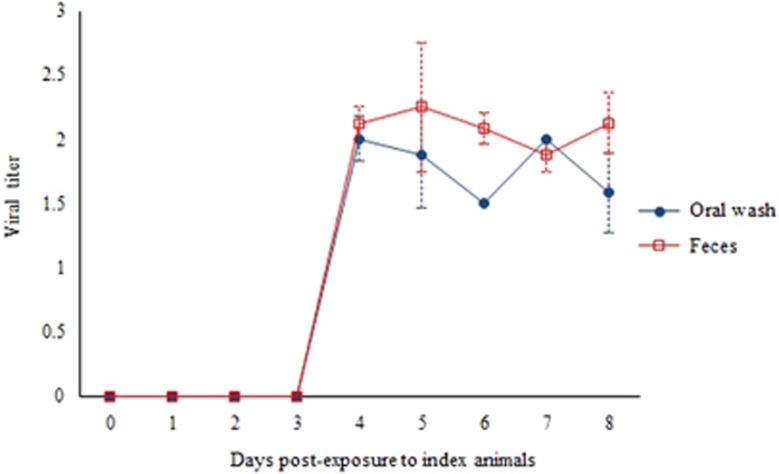
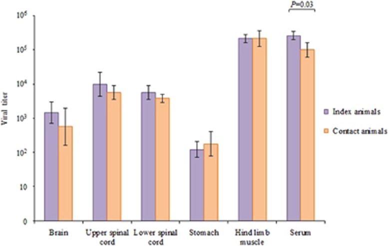
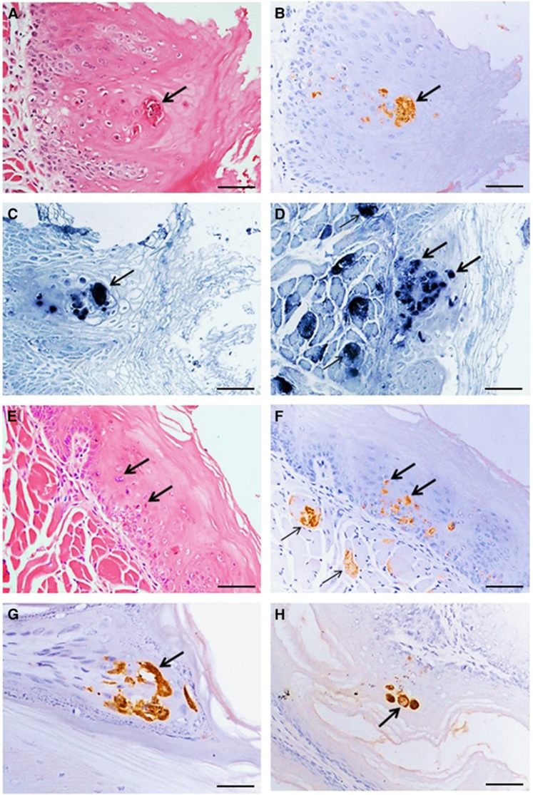
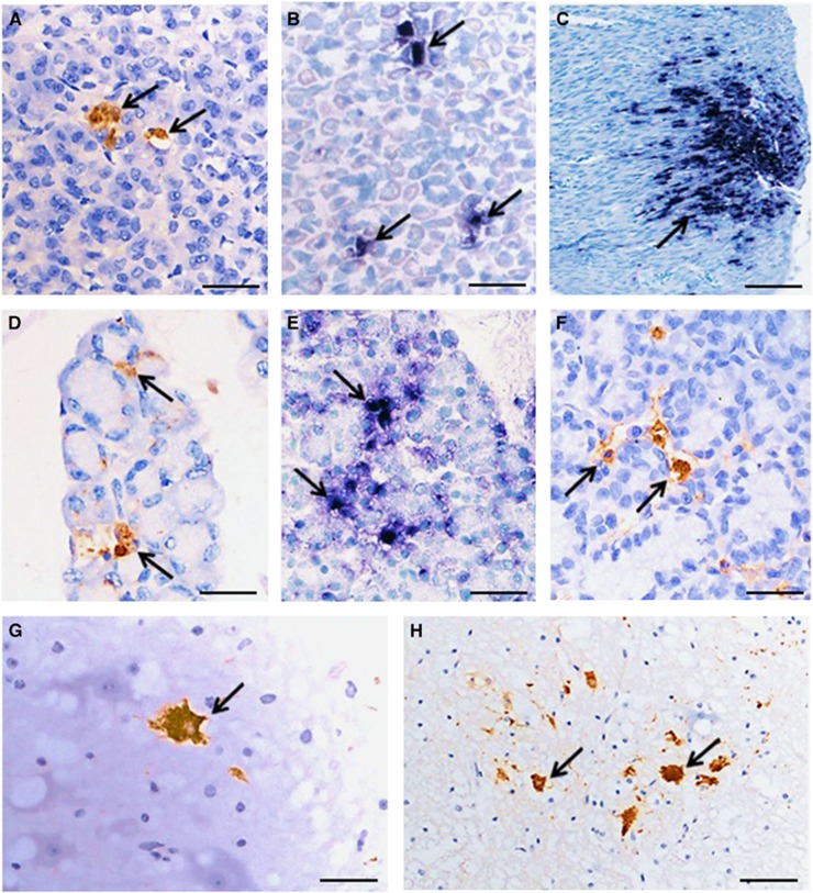
Enterovirus A71 (EV-A71) causes hand-foot-and-mouth disease (HFMD), which may be complicated by fatal encephalomyelitis. Although fecal-oral or oral-oral routes are important in person-to-person transmission, how viral shedding and exposure may predispose individuals to infection remains unknown. We investigated person-to-person transmission by using a model of HFMD and encephalomyelitis based on EV-A71 oral infection of 2-week-old hamsters. Animals (index animals) infected with 104 50% cell culture infective doses of virus uniformly developed severe disease four days post-infection (dpi), whereas littermate contacts developed severe disease after six to seven days of exposure to index animals. Virus was detected in oral washes and feces at 3-4 dpi in index animals and at three to eight days after exposure to index animals in littermate contact animals. In a second experiment, non-littermate contact animals exposed for 8 or 12 h to index animals developed the disease six and four days post-exposure, respectively. Tissues from killed index and contact animals, studied by light microscopy, immunohistochemistry and in situ hybridization, exhibited mild inflammatory lesions and/or viral antigens/RNA in the squamous epithelia of the oral cavity, tongue, paws, skin, esophagus, gastric epithelium, salivary glands, lacrimal glands, central nervous system neurons, muscles (skeletal, cardiac and smooth muscles) and liver. Orally shed viruses were probably derived from infected oral mucosa and salivary glands, whereas fecal viruses may have derived from these sites as well as from esophageal and gastric epithelia. Asymptomatic seroconversion in exposed mother hamsters was demonstrated. Our hamster model should be useful in studying person-to-person EV-A71 transmission and how drugs and vaccines may interrupt transmission.


Figures

Figure 1
Figure 1
Oral wash and fecal viral titers in index and littermate contact animals (n=4 each) at four days post-infection and eight days post-exposure, respectively, from experiment 1 (A). Oral wash and fecal viral titers in index animals and non-littermate contact animals (n=4 each) in the 12 h exposure group in experiment 2 (B). Viral titer is expressed as CCID50/mL±standard error of mean per 10% suspension. There were no significant differences between titers; all P values were >0.05.
Figure 2
Figure 2
Growth curve of viruses isolated without passage from oral washes and feces of contact animals from experiment 1. Virus titer is expressed as CCID50/mL±standard error of mean per of 10% suspension of oral samples and 10% (wt/vol) tissue homogenates of fecal samples.
Figure 3
Figure 3
Viral titers from various tissues of index and littermate contact animals (n=3 each) in experiment 1 at 4 days post-infection and 8 days post-exposure, respectively. Viral titer is expressed as the mean CCID50/mL±sem per 10% tissue homogenate. Viral titer in sera is expressed as CCID50/mL±sem per 10% dilution. There were no significant differences between tissue viral titers; P values were >0.05, with the exception of sera (P=0.03).
Figure 4
Figure 4
Graphs of ‘survival’ or period before killing of three groups of littermate contact animals (n=4 each group) from experiment 2 with 4, 8 and 12 h exposures to infected index animals.
Figure 5
Figure 5
Pathological findings in squamous cells and skeletal muscles in littermate contact hamsters at 8 days post-exposure (experiment 1). Squamous epithelial cell necrosis in the oral mucosa (A); localization of viral antigens (B) and viral RNA (C) in the same lesion in adjacent tissue sections. A mildly inflamed lesion in tongue squamous epithelial cells (E) has viral antigens in the same lesion (F) and skeletal muscle fibers (F) in adjacent tissue sections, as well as viral RNA in the same tongue squamous lesion (D) and skeletal muscle fibers (D). Viral antigens in squamous cells in paw epidermis (G) and esophageal mucosa (H). Stains: hematoxylin and eosin (A,E) immunohistochemistry with 3, 3’ diaminobenzidinetetrahydrochloride chromogen/hematoxylin (B,F,G,H) and in situ hybridization with nitroblue tetrazolium/5-bromo-4-chloro-3-indolyl phosphate/hematoxylin (C, D). Original magnification: 20x objective (A–F,H), × 40 objective (G). Scale bars: 30 μm (A–F,H), 15 μm (G).
Figure 6
Figure 6
Pathological findings in the orodigestive tract and central nervous system in littermate contact hamsters at 8 days post-exposure (experiment 1). Viral antigens (A) and viral RNA (B) in gastric mucosal epithelium, and viral RNA in gastric smooth muscle (C). Viral antigens (D) and viral RNA (E) were detected in salivary gland acinar cells. Viral antigens in lacrimal gland acinar cells (F), brainstem neurons (G) and spinal cord anterior horn cells (H) were also detected. Stains: Immunohistochemistry with 3, 3’ diaminobenzidinetetrahydrochloride chromogen/hematoxylin (A,D,F–H) and in situ hybridization with nitroblue tetrazolium/5-bromo-4-chloro-3-indolyl phosphate/hematoxylin (B,C,E). Original magnification: 10x objective (C), × 20 objective (H), and × 40 objective (A,B,D–G). Scale bars: 50 μm (C), 30 μm (H), and 15 μm (A,B,D–G).

Similar articles

Cited by

References

    1. Knipe DM, Howley PM. Fields Virology. In: Griffin DE et al (eds). Enteroviruses: Polioviruses, Coxsackieviruses, Echoviruses, and Newer Enteroviruses. 4th ed. USA: Lippincott Williams & Wilkins. 2001, 723–775.
    1. Yip CCY, Lau SKP, Zhou B et al. Emergence of enterovirus 71 “double-recombinant” strains belonging to a novel genotype D originating from southern China: first evidence for combination of intratypic and intertypic recombination events in EV71. Arch Virol 2010; 155: 1413–1424. - PMC - PubMed
    1. Ho M, Chen ER, Hsu KH et al. An epidemic of enterovirus 71 infection in Taiwan. N Engl J Med 1999; 341: 929–935. - PubMed
    1. Ooi MH, Wong SC, Podin Y et al. Human enterovirus 71 disease in Sarawak, Malaysia: a prospective clinical, virological, and molecular epidemiological study. Clin Infect Dis 2007; 44: 646–656. - PubMed
    1. Mcminn PC. Enterovirus 71 in the Asia-Pacific region: an emerging cause of acute neurological disease in young children. Neurol J Southeast Asia 2003; 8: 57–63.

MeSH terms

LinkOut - more resources