Review
. 2017:2017:6501046.
doi: 10.1155/2017/6501046. Epub 2017 Jun 18.

Measurement and Clinical Significance of Biomarkers of Oxidative Stress in Humans

Affiliations
Review

Measurement and Clinical Significance of Biomarkers of Oxidative Stress in Humans

Ilaria Marrocco et al. Oxid Med Cell Longev. 2017.

Abstract

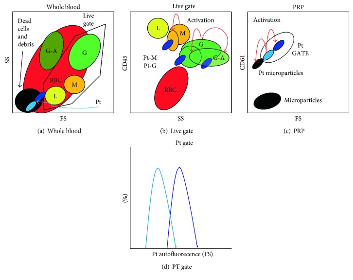
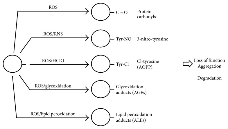
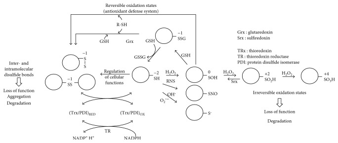
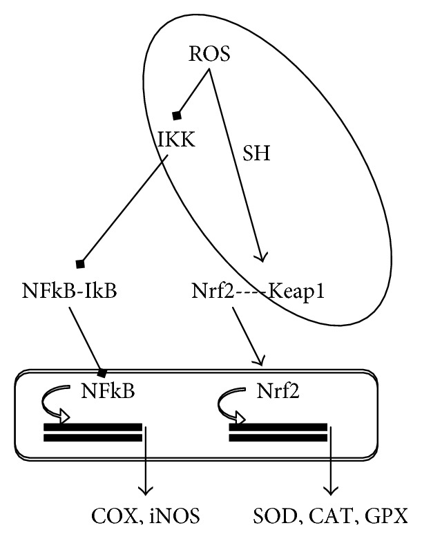
Oxidative stress is the result of the imbalance between reactive oxygen species (ROS) formation and enzymatic and nonenzymatic antioxidants. Biomarkers of oxidative stress are relevant in the evaluation of the disease status and of the health-enhancing effects of antioxidants. We aim to discuss the major methodological bias of methods used for the evaluation of oxidative stress in humans. There is a lack of consensus concerning the validation, standardization, and reproducibility of methods for the measurement of the following: (1) ROS in leukocytes and platelets by flow cytometry, (2) markers based on ROS-induced modifications of lipids, DNA, and proteins, (3) enzymatic players of redox status, and (4) total antioxidant capacity of human body fluids. It has been suggested that the bias of each method could be overcome by using indexes of oxidative stress that include more than one marker. However, the choice of the markers considered in the global index should be dictated by the aim of the study and its design, as well as by the clinical relevance in the selected subjects. In conclusion, the clinical significance of biomarkers of oxidative stress in humans must come from a critical analysis of the markers that should give an overall index of redox status in particular conditions.


Figures

Figure 1
Figure 1
Gating strategies in the measure of free-radical production by flow cytometry. Different leukocytes populations (lymphocytes: L, monocytes: M, and granulocytes: G) in whole blood can be identified by CD45 (b) in the live gate assigned in the forward scatter (FS) and side scatter (SS) dot plot (a) by excluding dead cells and debris. Red blood cells (RBC) can be excluded as CD45 negative (b). Platelets (Pt) can be identified by CD61 in platelet-rich plasma (PRP) (c). In activated samples, platelet microparticles (c) and leukocyte-platelet aggregates (b: Pt-G and Pt-M) are formed and Pt-G are more prone to apoptosis (G-A). After platelet activation, FS increases due to platelet aggregation inducing an increase in autofluorescence (d).
Figure 2
Figure 2
Irreversible oxidative modifications of proteins. AGEs: advanced glycation end products; ALEs: advanced peroxidation end products; AOPP: advanced oxidation protein products; HClO: hypochlorous acid; RNS: reactive nitrogen species; ROS reactive oxygen species.
Figure 3
Figure 3
Reversible oxidation of protein cysteine residues. GSH: glutathione; H2O2: hydrogen peroxide; O2•−: superoxide; RNS: reactive nitrogen species; RS: sulfur atom; −SO2H: sulfinic acid; −SO3H: sulfonic acid; −SOH: sulfenic acid.
Figure 4
Figure 4
Cysteine-regulated gene expression. CAT catalase; COX: cyclooxygenase; GPX: glutathione peroxidase; IKK: Iκ kinases; iNOS: inducible nitric oxide synthase; Keap1: Kelch-like ECH-associating protein 1; Nfr2: nuclear factor-erythroid 2-related factor 2; NF-kB: nuclear factor kappa-light-chain-enhancer of activated B cells; ROS: reactive oxygen species; SH: thiol; SOD: superoxide dismutase.
Figure 5
Figure 5
ROS generating enzymes. H2O2: hydrogen peroxide; HClO: hypochlorous acid; MPO: myeloperoxidase; NOS: NO synthase; NOX: NADPH oxidase; O2•−: superoxide; XO: xanthine oxidase.
Figure 6
Figure 6
Antioxidant enzymes. CAT: catalase; GPX: glutathione peroxidase; GR: glutathione reductase; H2O2: hydrogen peroxide; O2•−: superoxide; SOD: superoxide dismutase.

Similar articles

Cited by

References

    1. Valko M., Leibfritz D., Moncol J., Cronin M. T., Mazur M., Telser J. Free radicals and antioxidants in normal physiological functions and human disease. The International Journal of Biochemistry & Cell Biology. 2007;39(1):44–84. doi: 10.1016/j.biocel.2006.07.001. - DOI - PubMed
    1. Bickers D. R., Athar M. Oxidative stress in the pathogenesis of skin disease. The Journal of Investigative Dermatology. 2006;126(12):2565–2575. doi: 10.1038/sj.jid.5700340. - DOI - PubMed
    1. Franco R., Sanchez-Olea R., Reyes-Reyes E. M., Panayiotidis M. I. Environmental toxicity, oxidative stress and apoptosis: menage a trois. Mutation Research. 2009;674(1-2):3–22. - PubMed
    1. Hodjat M., Rezvanfar M. A., Abdollahi M. A systematic review on the role of environmental toxicants in stem cells aging. Food and Chemical Toxicology. 2015;86:298–308. doi: 10.1016/j.fct.2015.11.002. - DOI - PubMed
    1. Negre-Salvayre A., Auge N., Ayala V., et al. Pathological aspects of lipid peroxidation. Free Radical Research. 2010;44(10):1125–1171. doi: 10.3109/10715762.2010.498478. - DOI - PubMed