

The knee meniscus: management of traumatic tears and degenerative lesions
- PMID: 28698804
- PMCID: PMC5489759
- DOI: 10.1302/2058-5241.2.160056
The knee meniscus: management of traumatic tears and degenerative lesions
Abstract
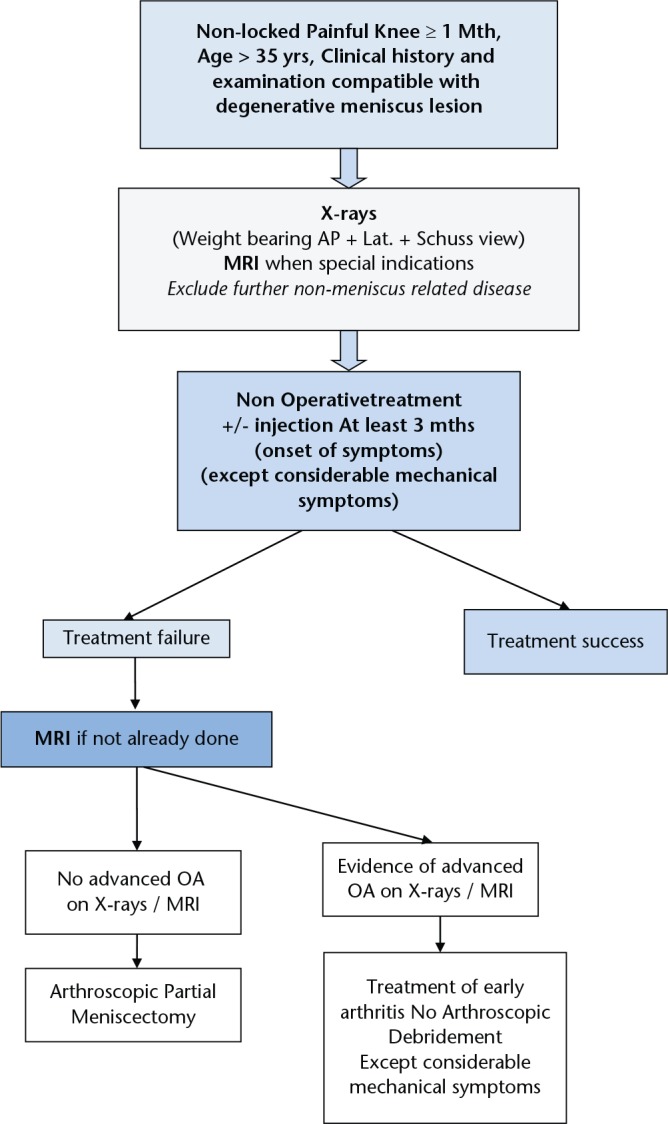
Meniscectomy is one of the most popular orthopaedic procedures, but long-term results are not entirely satisfactory and the concept of meniscal preservation has therefore progressed over the years. However, the meniscectomy rate remains too high even though robust scientific publications indicate the value of meniscal repair or non-removal in traumatic tears and non-operative treatment rather than meniscectomy in degenerative meniscal lesionsIn traumatic tears, the first-line choice is repair or non-removal. Longitudinal vertical tears are a proper indication for repair, especially in the red-white or red-red zones. Success rate is high and cartilage preservation has been proven. Non-removal can be discussed for stable asymptomatic lateral meniscal tears in conjunction with anterior cruciate ligament (ACL) reconstruction. Extended indications are now recommended for some specific conditions: horizontal cleavage tears in young athletes, hidden posterior capsulo-meniscal tears in ACL injuries, radial tears and root tears.Degenerative meniscal lesions are very common findings which can be considered as an early stage of osteoarthritis in middle-aged patients. Recent randomised studies found that arthroscopic partial meniscectomy (APM) has no superiority over non-operative treatment. Thus, non-operative treatment should be the first-line choice and APM should be considered in case of failure: three months has been accepted as a threshold in the ESSKA Meniscus Consensus Project presented in 2016. Earlier indications may be proposed in cases with considerable mechanical symptoms.The main message remains: save the meniscus! Cite this article: EFORT Open Rev 2017;2. DOI: 10.1302/2058-5241.2.160056. Originally published online at www.efortopenreviews.org.
Keywords: consensus; degenerative meniscal lesions; guidelines; knee; meniscectomy; meniscus; meniscus repair.
Conflict of interest statement
ICMJE Conflict of Interest Statement: PB: Occasional Education Consultant for Zimmer/Biomet, and Smith & Nephew. Editor in Chief of Orthopaedics and Traumatology: Surgery and Research RB: Deputy Editor of Knee Surgery Sports Traumatology and Arthroscopy, Education Consultant for Mathys, Wolf SK: web-editor of Knee Surgery Sports Traumatology and Arthroscopy MO: No conflict with this article NP: Occasional Education Consultant for Smith & Nephew, Zimmer/Biomet
Figures
References
-
- Smillie IS. The current pattern of internal derangements of the knee joint relative to the menisci. Clin Orthop Relat Res 1967;51:117-122 - PubMed
-
- Arnoczky SP, Warren RF. The microvasculature of the meniscus and its response to injury. An experimental study in the dog. Am J Sports Med 1983;11:131-141. - PubMed
-
- No authors listed. Agence Technique de l’Information sur l’Hospitalisation (ATIH). http://www.atih.sante.fr/mco/presentation?secteur=MCO (date last accessed 4 January 2017). (In French)
-
- Beaufils P, Hulet C, Dhénain M, et al. Clinical practice guidelines for the management of meniscal lesions and isolated lesions of the anterior cruciate ligament of the knee in adults. Orthop Traumatol Surg Res 2009;95:437-442. - PubMed
LinkOut - more resources
Full Text Sources
Other Literature Sources
