

Circadian Rhythms in Fear Conditioning: An Overview of Behavioral, Brain System, and Molecular Interactions
- PMID: 28698810
- PMCID: PMC5494081
- DOI: 10.1155/2017/3750307
Circadian Rhythms in Fear Conditioning: An Overview of Behavioral, Brain System, and Molecular Interactions
Abstract
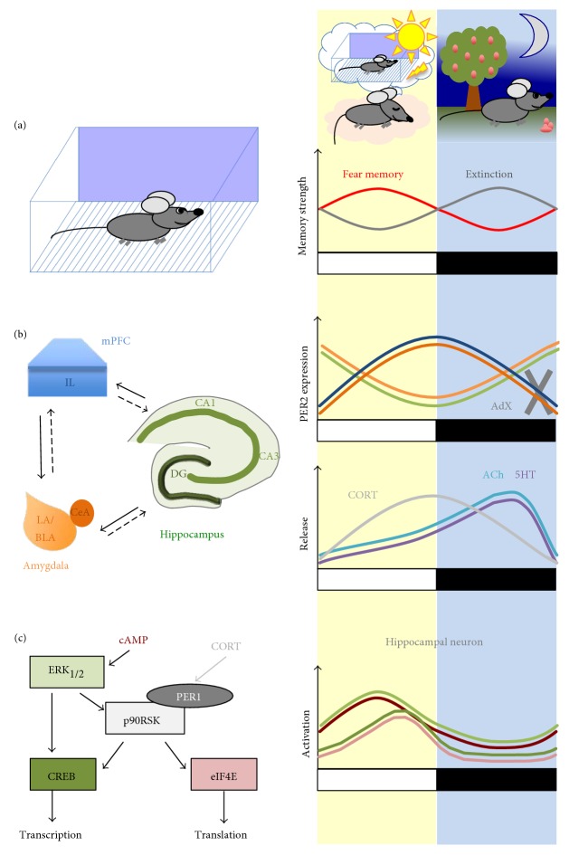
The formation of fear memories is a powerful and highly evolutionary conserved mechanism that serves the behavioral adaptation to environmental threats. Accordingly, classical fear conditioning paradigms have been employed to investigate fundamental molecular processes of memory formation. Evidence suggests that a circadian regulation mechanism allows for a timestamping of such fear memories and controlling memory salience during both their acquisition and their modification after retrieval. These mechanisms include an expression of molecular clocks in neurons of the amygdala, hippocampus, and medial prefrontal cortex and their tight interaction with the intracellular signaling pathways that mediate neural plasticity and information storage. The cellular activities are coordinated across different brain regions and neural circuits through the release of glucocorticoids and neuromodulators such as acetylcholine, which integrate circadian and memory-related activation. Disturbance of this interplay by circadian phase shifts or traumatic experience appears to be an important factor in the development of stress-related psychopathology, considering these circadian components are of critical importance for optimizing therapeutic approaches to these disorders.
Figures
Similar articles
-
The GABA-synthetic enzyme GAD65 controls circadian activation of conditioned fear pathways.Behav Brain Res. 2014 Mar 1;260:92-100. doi: 10.1016/j.bbr.2013.11.042. Epub 2013 Dec 1. Behav Brain Res. 2014. PMID: 24300892
-
Building and burying fear memories in the brain.Neuroscientist. 2005 Feb;11(1):89-99. doi: 10.1177/1073858404269232. Neuroscientist. 2005. PMID: 15632281 Review.
-
Mesolimbic dopaminergic pathways in fear conditioning.Prog Neurobiol. 2004 Dec;74(5):301-20. doi: 10.1016/j.pneurobio.2004.09.004. Prog Neurobiol. 2004. PMID: 15582224 Review.
-
Encoding of fear learning and memory in distributed neuronal circuits.Nat Neurosci. 2014 Dec;17(12):1644-54. doi: 10.1038/nn.3869. Epub 2014 Nov 21. Nat Neurosci. 2014. PMID: 25413091 Review.
-
Cholinergic Signaling Controls Conditioned Fear Behaviors and Enhances Plasticity of Cortical-Amygdala Circuits.Neuron. 2016 Jun 1;90(5):1057-70. doi: 10.1016/j.neuron.2016.04.028. Epub 2016 May 5. Neuron. 2016. PMID: 27161525 Free PMC article.
Cited by
-
Emotionally clocked out: cell-type specific regulation of mood and anxiety by the circadian clock system in the brain.Front Mol Neurosci. 2023 Jun 27;16:1188184. doi: 10.3389/fnmol.2023.1188184. eCollection 2023. Front Mol Neurosci. 2023. PMID: 37441675 Free PMC article. Review.
-
Tracking conditioned fear in pair-housed mice using deep learning and real-time cue delivery.Neurobiol Stress. 2025 Jun 19;37:100742. doi: 10.1016/j.ynstr.2025.100742. eCollection 2025 Jul. Neurobiol Stress. 2025. PMID: 40678084 Free PMC article.
-
Differential Expression of Circadian Clock Genes in the Bovine Neuroendocrine Adrenal System.Genes (Basel). 2023 Nov 15;14(11):2082. doi: 10.3390/genes14112082. Genes (Basel). 2023. PMID: 38003025 Free PMC article.
-
Midday meals do not impair mouse memory.Sci Rep. 2018 Nov 19;8(1):17013. doi: 10.1038/s41598-018-35427-y. Sci Rep. 2018. PMID: 30451946 Free PMC article.
-
A hypothalamic circuit for the circadian control of aggression.Nat Neurosci. 2018 May;21(5):717-724. doi: 10.1038/s41593-018-0126-0. Epub 2018 Apr 9. Nat Neurosci. 2018. PMID: 29632359 Free PMC article.
References
Publication types
MeSH terms
LinkOut - more resources
Full Text Sources
Other Literature Sources
