Comparison of the characteristics of two hemoglobin variants, Hb D-Iran and Hb E, eluting in the Hb A2 window
- PMID: 28698850
- PMCID: PMC5503891
- DOI: 10.5045/br.2017.52.2.130
Comparison of the characteristics of two hemoglobin variants, Hb D-Iran and Hb E, eluting in the Hb A2 window
Abstract
Background: Cation exchange-high performance liquid chromatography (CE-HPLC) is most commonly used to evaluate hemoglobin (Hb) variants, which elute in the Hb A2 window. This study aimed to assess prevalence of an uncommon Hb variant, Hb D-Iran, and compare its red cell parameters and peak characteristics with those of Hb E that commonly elutes in the Hb A2 window.
Methods: Generally, we assess abnormal Hb using CE-HPLC as the primary technique along with alkaline and acid electrophoresis. All cases with Hb A2 window >9%, as assessed by CE-HPLCs during 2009-2013, were selected.
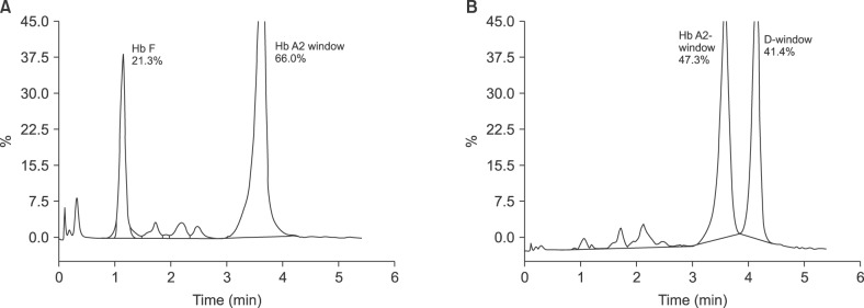
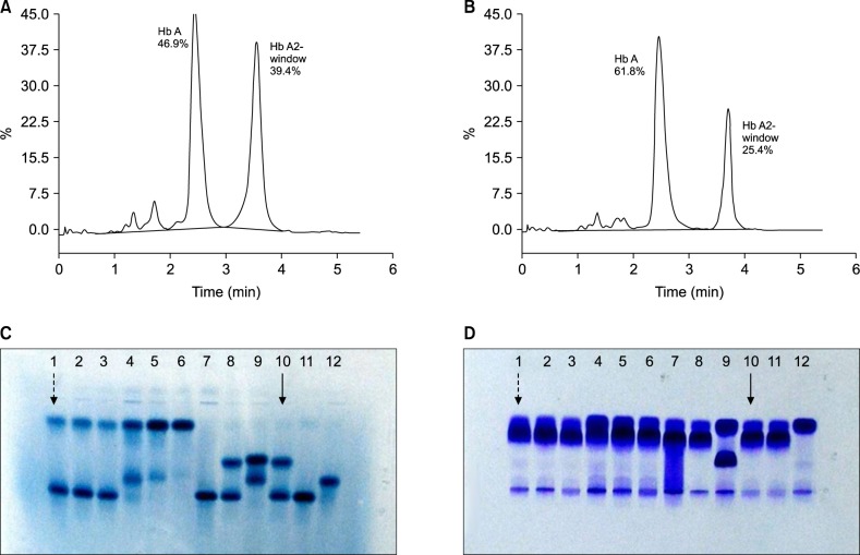
Results: Twenty-nine cases with Hb D-Iran variant were identified-25 heterozygous, 2 homozygous, 1 compound heterozygous Hb D-Iran/β-thalassemia, and 1 Hb D-Iran/Hb D-Punjab. Overall prevalence of Hb D-Iran was 0.23%. Compared to patients with Hb E, those with Hb D-Iran had significantly higher Hb (12.1 vs. 11.3 g/dL, P=0.03), MCV (82.4 vs. 76.4 fL, P=0.0044), MCH (27.9 vs. 25.45 pg, P =0.0006), and MCHC (33.9 vs. 33.3 g/dL, P=0.0005). Amount of abnormal Hb (40.7 vs. 26.4%, P=0.0001) was significantly higher while retention time (3.56 vs. 3.70 min, P=0.0001) was significantly lower in Hb D-Iran than in Hb E.
Conclusion: Hb D-Iran peak can be easily missed if area and retention time of the Hb A2 window are not carefully analyzed. To distinguish between variants, careful analysis of peak area and retention time is sufficient in most cases and may be further confirmed by the second technique-alkaline electrophoresis.
Keywords: CE-HPLC; Hb A2 window; Hb D-Iran; Hb E.
Conflict of interest statement
Authors' Disclosures of Potential Conflicts of Interest: No potential conflicts of interest relevant to this article were reported.
Figures
Similar articles
-
Compound heterozygous hemoglobin d-punjab/hemoglobin d-iran: a novel hemoglobinopathy.Indian J Hematol Blood Transfus. 2014 Sep;30(Suppl 1):409-12. doi: 10.1007/s12288-014-0441-x. Epub 2014 Aug 8. Indian J Hematol Blood Transfus. 2014. PMID: 25332633 Free PMC article.
-
Comparison of Two High-Pressure Liquid Chromatography Instruments Bio-Rad Variant-II and Tosoh HLC-723G11 in the Evaluation of Hemoglobinopathies.Indian J Hematol Blood Transfus. 2020 Oct;36(4):725-732. doi: 10.1007/s12288-020-01298-5. Epub 2020 Jun 19. Indian J Hematol Blood Transfus. 2020. PMID: 33100717 Free PMC article.
-
Diagnosis of a rare double heterozygous Hb D Punjab/Hb Q India hemoglobinopathy using Sebia capillary zone electrophoresis.Indian J Pathol Microbiol. 2014 Oct-Dec;57(4):626-8. doi: 10.4103/0377-4929.142709. Indian J Pathol Microbiol. 2014. PMID: 25308024
-
Detection of Hemoglobin Constant Spring: A Comparison of Capillary Electrophoresis Versus High-Performance Liquid Chromatography.Cureus. 2024 Aug 19;16(8):e67228. doi: 10.7759/cureus.67228. eCollection 2024 Aug. Cureus. 2024. PMID: 39295657 Free PMC article.
-
Update in Laboratory Diagnosis of Thalassemia.Front Mol Biosci. 2020 May 27;7:74. doi: 10.3389/fmolb.2020.00074. eCollection 2020. Front Mol Biosci. 2020. PMID: 32671092 Free PMC article. Review.
Cited by
-
Epidemiologic profile of hemoglobinopathies in Benin.Hematol Transfus Cell Ther. 2024 Dec;46 Suppl 6(Suppl 6):S257-S262. doi: 10.1016/j.htct.2024.07.008. Epub 2024 Oct 8. Hematol Transfus Cell Ther. 2024. PMID: 39426879 Free PMC article.
-
Fetal heterozygosity for both Hb G-Hsi-Tsou and beta thalassemia: A case report.Case Rep Womens Health. 2020 Oct 22;28:e00265. doi: 10.1016/j.crwh.2020.e00265. eCollection 2020 Oct. Case Rep Womens Health. 2020. PMID: 33163367 Free PMC article.
-
Case report-unveiling a case of hemoglobin D-Punjab variant with iron deficiency anemia in Sindh (Pakistan).SAGE Open Med Case Rep. 2024 Aug 16;12:2050313X241272516. doi: 10.1177/2050313X241272516. eCollection 2024. SAGE Open Med Case Rep. 2024. PMID: 39161918 Free PMC article.
References
-
- Rahbar S. Haemoglobin D Iran: β222 glutamic acid leads to glutamine (B4) Br J Haematol. 1973;24:31–35. - PubMed
-
- Colah RB, Surve R, Sawant P, et al. HPLC studies in hemoglobinopathies. Indian J Pediatr. 2007;74:657–662. - PubMed
-
- Olivieri NF, Pakbaz Z, Vichinsky E. HbE/β-thalassemia: basis of marked clinical diversity. Hematol Oncol Clin North Am. 2010;24:1055–1070. - PubMed
LinkOut - more resources
Full Text Sources
Other Literature Sources