

Comparative ultrastructure of fruit plastids in three genetically diverse genotypes of apple (Malus × domestica Borkh.) during development
- PMID: 28698906
- PMCID: PMC5693628
- DOI: 10.1007/s00299-017-2179-z
Comparative ultrastructure of fruit plastids in three genetically diverse genotypes of apple (Malus × domestica Borkh.) during development
Abstract
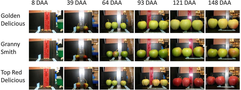
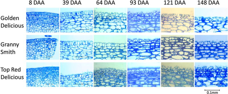
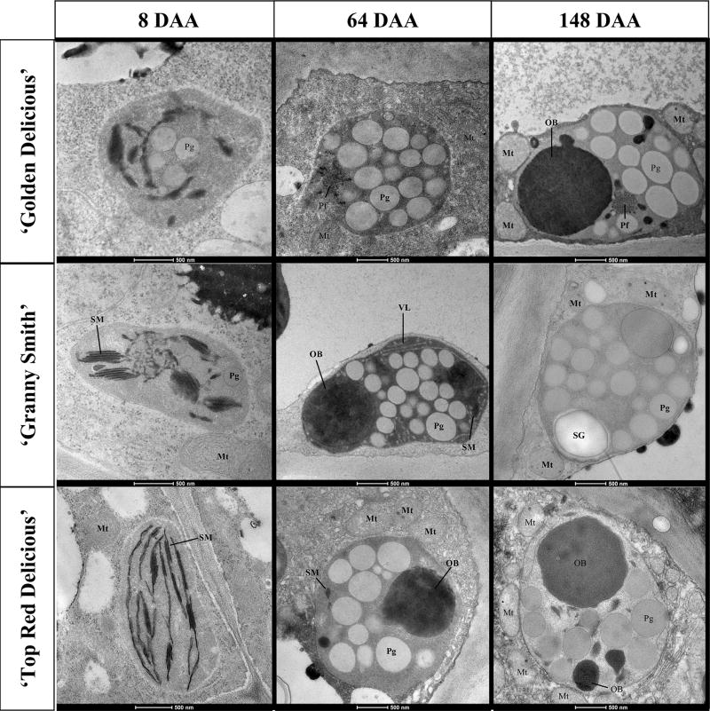
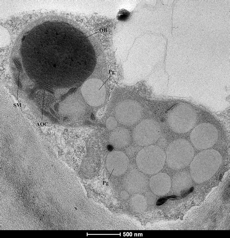
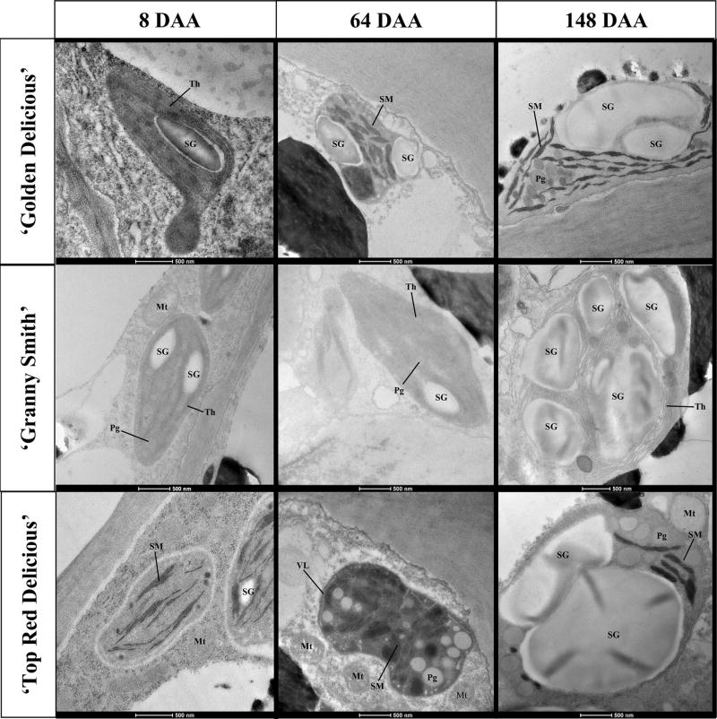
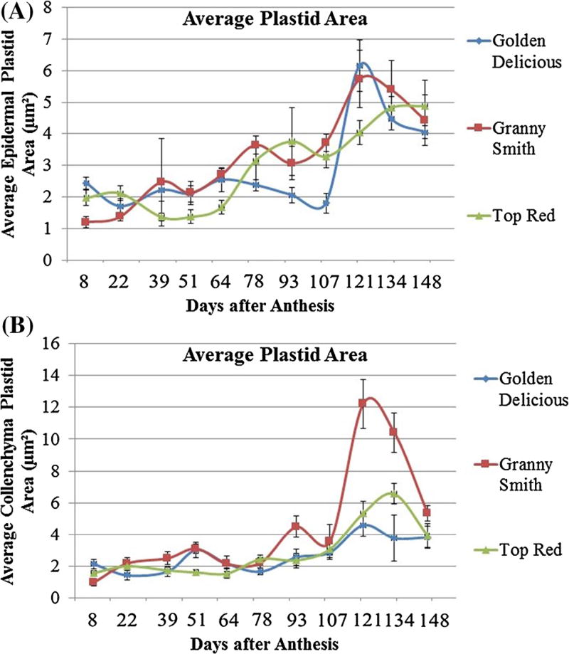
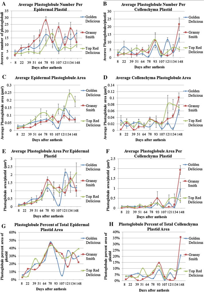
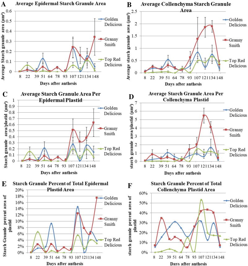
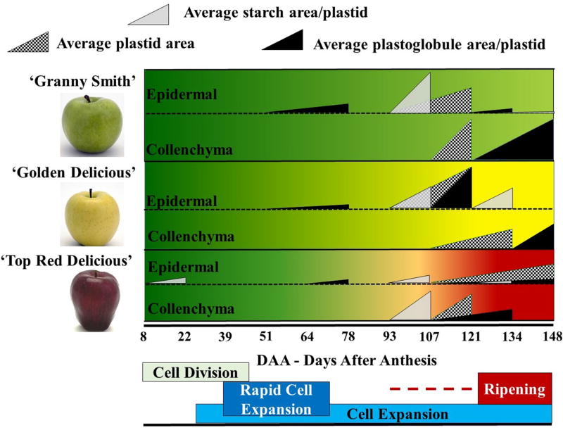
Comparative ultrastructural developmental time-course analysis has identified discrete stages at which the fruit plastids undergo structural and consequently functional transitions to facilitate subsequent development-guided understanding of the complex plastid biology. Plastids are the defining organelle for a plant cell and are critical for myriad metabolic functions. The role of leaf plastid, chloroplast, is extensively documented; however, fruit plastids-chromoplasts-are poorly understood, especially in the context of the diverse metabolic processes operating in these diverse plant organs. Recently, in a comparative study of the predicted plastid-targeted proteomes across seven plant species, we reported that each plant species is predicted to harbor a unique set of plastid-targeted proteins. However, the temporal and developmental context of these processes remains unknown. In this study, an ultrastructural analysis approach was used to characterize fruit plastids in the epidermal and collenchymal cell layers at 11 developmental timepoints in three genotypes of apple (Malus × domestica Borkh.): chlorophyll-predominant 'Granny Smith', carotenoid-predominant 'Golden Delicious', and anthocyanin-predominant 'Top Red Delicious'. Plastids transitioned from a proplastid-like plastid to a chromoplast-like plastid in epidermis cells, while in the collenchyma cells, they transitioned from a chloroplast-like plastid to a chloro-chromo-amyloplast plastid. Plastids in the collenchyma cells of the three genotypes demonstrated a diverse array of structures and features. This study enabled the identification of discrete developmental stages during which specific functions are most likely being performed by the plastids as indicated by accumulation of plastoglobuli, starch granules, and other sub-organeller structures. Information regarding the metabolically active developmental stages is expected to facilitate biologically relevant omics studies to unravel the complex biochemistry of plastids in perennial non-model systems.
Keywords: Chloroplast; Chromoplast; Malus × domestica Borkh.; Plastid transition; Rosaceae.
Conflict of interest statement
Figures
Similar articles
-
The effect of promoter methylation on MdMYB1 expression determines the level of anthocyanin accumulation in skins of two non-red apple cultivars.BMC Plant Biol. 2018 Jun 5;18(1):108. doi: 10.1186/s12870-018-1320-7. BMC Plant Biol. 2018. PMID: 29871614 Free PMC article.
-
Metabolic and gene expression analysis of apple (Malus x domestica) carotenogenesis.J Exp Bot. 2012 Jul;63(12):4497-511. doi: 10.1093/jxb/ers134. Epub 2012 Jun 19. J Exp Bot. 2012. PMID: 22717407 Free PMC article.
-
Comparative analysis of predicted plastid-targeted proteomes of sequenced higher plant genomes.PLoS One. 2014 Nov 13;9(11):e112870. doi: 10.1371/journal.pone.0112870. eCollection 2014. PLoS One. 2014. PMID: 25393533 Free PMC article.
-
Differentiation of chromoplasts and other plastids in plants.Plant Cell Rep. 2019 Jul;38(7):803-818. doi: 10.1007/s00299-019-02420-2. Epub 2019 May 11. Plant Cell Rep. 2019. PMID: 31079194 Free PMC article. Review.
-
Chromoplast differentiation: current status and perspectives.Plant Cell Physiol. 2010 Oct;51(10):1601-11. doi: 10.1093/pcp/pcq136. Epub 2010 Aug 27. Plant Cell Physiol. 2010. PMID: 20801922 Review.
Cited by
-
Non-aqueous fractionation revealed changing subcellular metabolite distribution during apple fruit development.Hortic Res. 2019 Aug 11;6:98. doi: 10.1038/s41438-019-0178-7. eCollection 2019. Hortic Res. 2019. PMID: 31666959 Free PMC article.
-
Genome-Scale Characterization of Predicted Plastid-Targeted Proteomes in Higher Plants.Sci Rep. 2020 May 19;10(1):8281. doi: 10.1038/s41598-020-64670-5. Sci Rep. 2020. PMID: 32427841 Free PMC article.
-
Carotenoid Biosynthesis and Plastid Development in Plants: The Role of Light.Int J Mol Sci. 2021 Jan 26;22(3):1184. doi: 10.3390/ijms22031184. Int J Mol Sci. 2021. PMID: 33530294 Free PMC article. Review.
-
Cutin Synthesis in Developing, Field-Grown Apple Fruit Examined by External Feeding of Labelled Precursors.Plants (Basel). 2021 Mar 5;10(3):497. doi: 10.3390/plants10030497. Plants (Basel). 2021. PMID: 33807966 Free PMC article.
-
Comparative transcriptomic and plastid development analysis sheds light on the differential carotenoid accumulation in kiwifruit flesh.Front Plant Sci. 2023 Aug 30;14:1213086. doi: 10.3389/fpls.2023.1213086. eCollection 2023. Front Plant Sci. 2023. PMID: 37711308 Free PMC article.
References
-
- Abramoff M, Magalhaes P, Ram S. Image processing with ImageJ. Biophoton Int. 2004;11:36–42.
-
- Alexander L, Grierson D. Ethylene biosynthesis and action in tomato: a model for climacteric fruit ripening. J Exp Bot. 2002;53:2039–2055. - PubMed
-
- Barsan C, Sanchez-Bel P, Rombaldi C, Egea I, Rossignol M, Kuntz M, Zouine M, Latche A, Bouzayen M, Pech JC. Characteristics of the tomato chromoplast revealed by proteomic analysis. J Exp Bot. 2010;61:2413–2431. - PubMed
-
- Bartley IM, Stoker PG, Martin ADE, Hatfield SGS, Knee M. Synthesis of aroma compounds by apples supplied with alcohols and methyl-esters of fatty-acids. J Sci Food Agric. 1985;36:567–574.
-
- Bizjak J, Mikulic-Petkovsek M, Stampar F, Veberic R. Changes in primary metabolites and polyphenols in the peel of “Braeburn” apples (Malus domestica Borkh.) during advanced maturation. J Agric Food Chem. 2013;61:10283–10292. - PubMed
Publication types
MeSH terms
Substances
Grants and funding
LinkOut - more resources
Full Text Sources
Other Literature Sources
