

Automated T2 relaxometry of the hippocampus for temporal lobe epilepsy
- PMID: 28699215
- PMCID: PMC5599984
- DOI: 10.1111/epi.13843
Automated T2 relaxometry of the hippocampus for temporal lobe epilepsy
Abstract
Objective: Hippocampal sclerosis (HS), the most common cause of refractory temporal lobe epilepsy, is associated with hippocampal volume loss and increased T2 signal. These can be identified on quantitative imaging with hippocampal volumetry and T2 relaxometry. Although hippocampal segmentation for volumetry has been automated, T2 relaxometry currently involves subjective and time-consuming manual delineation of regions of interest. In this work, we develop and validate an automated technique for hippocampal T2 relaxometry.
Methods: Fifty patients with unilateral or bilateral HS and 50 healthy controls underwent T1 -weighted and dual-echo fast recovery fast spin echo scans. Hippocampi were automatically segmented using a multi-atlas-based segmentation algorithm (STEPS) and a template database. Voxelwise T2 maps were determined using a monoexponential fit. The hippocampal segmentations were registered to the T2 maps and eroded to reduce partial volume effect. Voxels with T2 >170 msec excluded to minimize cerebrospinal fluid (CSF) contamination. Manual determination of T2 values was performed twice in each subject. Twenty controls underwent repeat scans to assess interscan reproducibility.
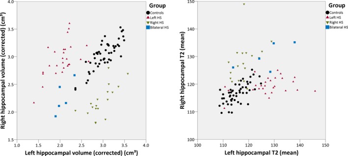
Results: Hippocampal T2 values were reliably determined using the automated method. There was a significant ipsilateral increase in T2 values in HS (p < 0.001), and a smaller but significant contralateral increase. The combination of hippocampal volumes and T2 values separated the groups well. There was a strong correlation between automated and manual methods for hippocampal T2 measurement (0.917 left, 0.896 right, both p < 0.001). Interscan reproducibility was superior for automated compared to manual measurements.
Significance: Automated hippocampal segmentation can be reliably extended to the determination of hippocampal T2 values, and a combination of hippocampal volumes and T2 values can separate subjects with HS from healthy controls. There is good agreement with manual measurements, and the technique is more reproducible on repeat scans than manual measurement. This protocol can be readily introduced into a clinical workflow for the assessment of patients with focal epilepsy.
Keywords: Hippocampus; Magnetic resonance imaging; T2 relaxometry; Temporal lobe epilepsy.
© 2017 The Authors. Epilepsia published by Wiley Periodicals, Inc. on behalf of International League Against Epilepsy.
Figures


Similar articles
-
Hippocampal profiling: Localized magnetic resonance imaging volumetry and T2 relaxometry for hippocampal sclerosis.Epilepsia. 2020 Feb;61(2):297-309. doi: 10.1111/epi.16416. Epub 2019 Dec 24. Epilepsia. 2020. PMID: 31872873 Free PMC article.
-
T2 relaxometry improves detection of non-sclerotic epileptogenic hippocampus.Epilepsy Res. 2016 Oct;126:1-9. doi: 10.1016/j.eplepsyres.2016.06.001. Epub 2016 Jun 25. Epilepsy Res. 2016. PMID: 27400070
-
Measurement of temporal lobe T2 relaxation times using a routine diagnostic MR imaging protocol in epilepsy.Epilepsy Res. 2002 Jan;48(1-2):131-42. doi: 10.1016/s0920-1211(01)00325-4. Epilepsy Res. 2002. PMID: 11823117
-
Advances in MRI-based diagnosis of temporal lobe epilepsy: Correlating hippocampal subfield volumes with histopathology.J Neuroimaging. 2024 Sep-Oct;34(5):515-526. doi: 10.1111/jon.13225. Epub 2024 Aug 2. J Neuroimaging. 2024. PMID: 39092876 Review.
-
Qualitative and quantitative imaging of the hippocampus in mesial temporal lobe epilepsy with hippocampal sclerosis.Neuroimaging Clin N Am. 2004 Aug;14(3):373-400, vii. doi: 10.1016/j.nic.2004.04.004. Neuroimaging Clin N Am. 2004. PMID: 15324854 Review.
Cited by
-
Combined quantitative T2 mapping and [18F]FDG PET could improve lateralization of mesial temporal lobe epilepsy.Eur Radiol. 2022 Sep;32(9):6108-6117. doi: 10.1007/s00330-022-08707-5. Epub 2022 Mar 28. Eur Radiol. 2022. PMID: 35347363 Free PMC article.
-
Histopathological Correlations of Qualitative and Quantitative Temporopolar MRI Analyses in Patients With Hippocampal Sclerosis.Front Neurol. 2021 Dec 24;12:801195. doi: 10.3389/fneur.2021.801195. eCollection 2021. Front Neurol. 2021. PMID: 35002940 Free PMC article.
-
MRI-T2 Relaxometry is Increased in Mild Traumatic Brain Injury: Indications of Acute Brain Abnormalities After Injury.J Neurosci Res. 2025 Apr;103(4):e70034. doi: 10.1002/jnr.70034. J Neurosci Res. 2025. PMID: 40178334 Free PMC article.
-
Enhancing Hippocampal Subfield Visualization Through Deep Learning Reconstructed MRI Scans.Diagnostics (Basel). 2025 Jun 16;15(12):1523. doi: 10.3390/diagnostics15121523. Diagnostics (Basel). 2025. PMID: 40564843 Free PMC article.
-
Magnetic resonance fingerprinting of temporal lobe white matter in mesial temporal lobe epilepsy.Ann Clin Transl Neurol. 2019 Sep;6(9):1639-1646. doi: 10.1002/acn3.50851. Epub 2019 Jul 30. Ann Clin Transl Neurol. 2019. PMID: 31359636 Free PMC article.
References
-
- Margerison JH, Corsellis JA. Epilepsy and the temporal lobes. A clinical, electroencephalographic and neuropathological study of the brain in epilepsy, with particular reference to the temporal lobes. Brain 1966;89:499–530. - PubMed
-
- Duncan JS, Sagar HJ. Seizure characteristics, pathology, and outcome after temporal lobectomy. Neurology 1987;37:405–409. - PubMed
-
- Jackson GD, Connelly A, Duncan JS, et al. Detection of hippocampal pathology in intractable partial epilepsy: increased sensitivity with quantitative magnetic resonance T2 relaxometry. Neurology 1993;43:1793–1799. - PubMed
Publication types
MeSH terms
Grants and funding
LinkOut - more resources
Full Text Sources
Other Literature Sources
