Review
. 2018 Mar;96(3):335-347.
doi: 10.1002/jnr.24116. Epub 2017 Jul 12.

Calcium dependence of spontaneous neurotransmitter release

Affiliations
Review

Calcium dependence of spontaneous neurotransmitter release

Courtney L Williams et al. J Neurosci Res. 2018 Mar.

Abstract

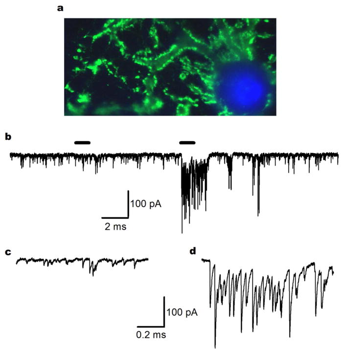
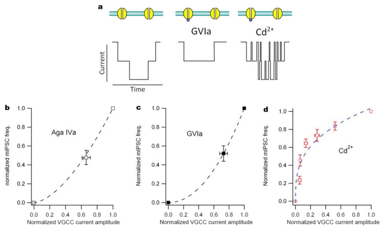
Spontaneous release of neurotransmitters is regulated by extracellular [Ca2+ ] and intracellular [Ca2+ ]. Curiously, some of the mechanisms of Ca2+ signaling at central synapses are different at excitatory and inhibitory synapses. While the stochastic activity of voltage-activated Ca2+ channels triggers a majority of spontaneous release at inhibitory synapses, this is not the case at excitatory nerve terminals. Ca2+ release from intracellular stores regulates spontaneous release at excitatory and inhibitory terminals, as do agonists of the Ca2+ -sensing receptor. Molecular machinery triggering spontaneous vesicle fusion may differ from that underlying evoked release and may be one of the sources of heterogeneity in release mechanisms.

Keywords: VGCC (voltage-gated calcium channels); calcium; minis; spontaneous release.


Figures

Figure 1
Figure 1
Random variability in the rate of spontaneous release. a Fluorescent micrograph labeled for synaptophysin (green) and DAPI (blue) illustrates that alarge number of nerve terminals may synapse onto a singleneocortical neuron. Spontaneous release will increase with the number of synaptic contacts, along with other factors including temperature. b Exemplary trace of miniature inhibitory post-synaptic currents (mIPSCs) from cortical cultured neurons recorded at −70 mV under previously described conditions (Williams et al. 2012). Horizontal bars above the trace indicate the traces used in c and d. The stochastic nature of spontaneous release results in a variable frequency and amplitude of events as shown. c Expanded time course from the trace in b to indicate basal mIPSC frequency. d Expanded timecourse from the trace in b to show a portion of the recording where a high frequency “burst” of mIPSCs was taking place.
Figure 2
Figure 2
Non-linear actions of VACC blockers on VACC currents and mIPSCs. a Cartoon of VACC current through two single channels indicating the effects of different types of channel blockers. The center illustration shows block by a selective “irreversible” blocker for N-type VACCs (GVIa, 1μM). The illustration on the right shows the result of blocking channels with Cd2+, which is nonselective and results in “flickery” block of the current (Lansman et al, 1986). b Plot of normalized mIPSC frequency versus normalized VGCC current amplitude in the presence and absence of a saturating dose of Aga Iva (300 nM), a selective P/Q-type channel antagonist. VACC currents were elicited by steps from −70 to −10 mV and mIPSC frequency was recorded at −70 mV, both in cultured neocortical neurons as previously described (Tsintsadze et al. 2017). Data are normalized average frequency (n=4) and average current (n=5) and represent the fraction the mIPSCs and VACC currents that are sensitive to a saturating dose of Cd2+ (300 μM). c Plot of normalized mIPSC frequency versus normalized VGCC current amplitude in the presence and absence of a saturating dose of GVIa (1μM), a selective N-type channel antagonist. Data are average frequency (n=9) and VACC current amplitude (n=5) recorded and normalized as for b. d Plot of normalized mIPSC frequency (n= 9) versus normalized VGCC current amplitude (n=13) in the presence of 0, 1, 3, 10, 30, 100, and 300 μM Cd2+. Recordings made as previously described(Tsintsadze et al. 2017). Note the differences in the shapes of the curves in b and c from the curve in d. If more than one channel contributes to spontaneous release, the relationship between release probability and the size of the Ca2+ current will be supralinear.
Figure 3
Figure 3
Single channel currents and Ca2+ domains. (Upper panel) Illustration of VACC current through two single channels in 1.1 and 6 mM [Ca2+]o. Note that the single channel current in 6 mM [Ca2+]o is approximately 3-fold larger than physiological [Ca2+]o (Weber et al. 2010). (Lower panel) Illustration showing the side view of the Ca2+ domain with 2 synaptic vesicles in 1.1 and 6 mM [Ca2+]o. With a higher [Ca2+]o, the Ca2+ domain may also increase 3-fold.
Figure 4
Figure 4
Spontaneous release is increased by external calcium in cultured neocortical neurons without MVR. a and g Exemplary mIPSC and mEPSCs, respectively, in 1.1 (top, black) and 6 (bottom, blue/red) mM [Ca2+]o. Recordings were made as reported previously (Vyleta and Smith 2011; Williams et al. 2012). b and h Histogram of inter-event interval (IEI) for mIPSCs or mEPSCs in 1.1 (black) and 6 (blue/red) mM [Ca2+]o from the same experiments in a and g. The signal-to-noise is reduced by plotting the square root of the ordinate and using logarithmic binning. Histograms are fit with double exponentials. c and i Histogram of amplitude of mIPSCs or mEPSCs in 1.1 (black) and 6 (blue/red) mM [Ca2+]o from the same experiments in a and g. dk Cumulative probability plots for IEI and amplitude of mIPSCs or mEPSCs in 1.1 (black) and 6 (blue/red) mM [Ca2+]o from the same experiments in a and g. f and l Average time constants (1st and 2nd Taus) from the double exponential fit of the IEI histograms for mIPSCs (n=9) and mEPSCs (n=8). Black bars indicate time constants in 1.1 [Ca2+]o and 6 blue/red bars represent time constants in 6 mM [Ca2+]o.

References

    1. Abenavoli A, Forti L, Bossi M, Bergamaschi A, Villa A, Malgaroli A. Multimodal quantal release at individual hippocampal synapses: evidence for no lateral inhibition. The Journal of neuroscience: the official journal of the Society for Neuroscience. 2002;22(15):6336–6346. - PMC - PubMed
    1. Ahmed MS, Siegelbaum SA. Recruitment of N-Type Ca(2+) channels during LTP enhances low release efficacy of hippocampal CA1 perforant path synapses. Neuron. 2009;63(3):372–385. - PMC - PubMed
    1. Andreae LC, Fredj NB, Burrone J. Independent vesicle pools underlie different modes of release during neuronal development. The Journal of neuroscience: the official journal of the Society for Neuroscience. 2012;32(5):1867–1874. - PMC - PubMed
    1. Augustine GJ, Charlton MP. Calcium dependence of presynaptic calcium current and post-synaptic response at the squid giant synapse. J Physiol. 1986;381(1):619–640. - PMC - PubMed
    1. Autry AE, Megumi A, Elena N, Na ES, Los MF, Peng-fei C, Kavalali ET, Monteggia LM. NMDA receptor blockade at rest triggers rapid behavioural antidepressant responses. Nature. 2011;475(7354):91–95. - PMC - PubMed

Publication types

MeSH terms