

Regulatory T cells in skin
- PMID: 28699278
- PMCID: PMC5629423
- DOI: 10.1111/imm.12791
Regulatory T cells in skin
Abstract
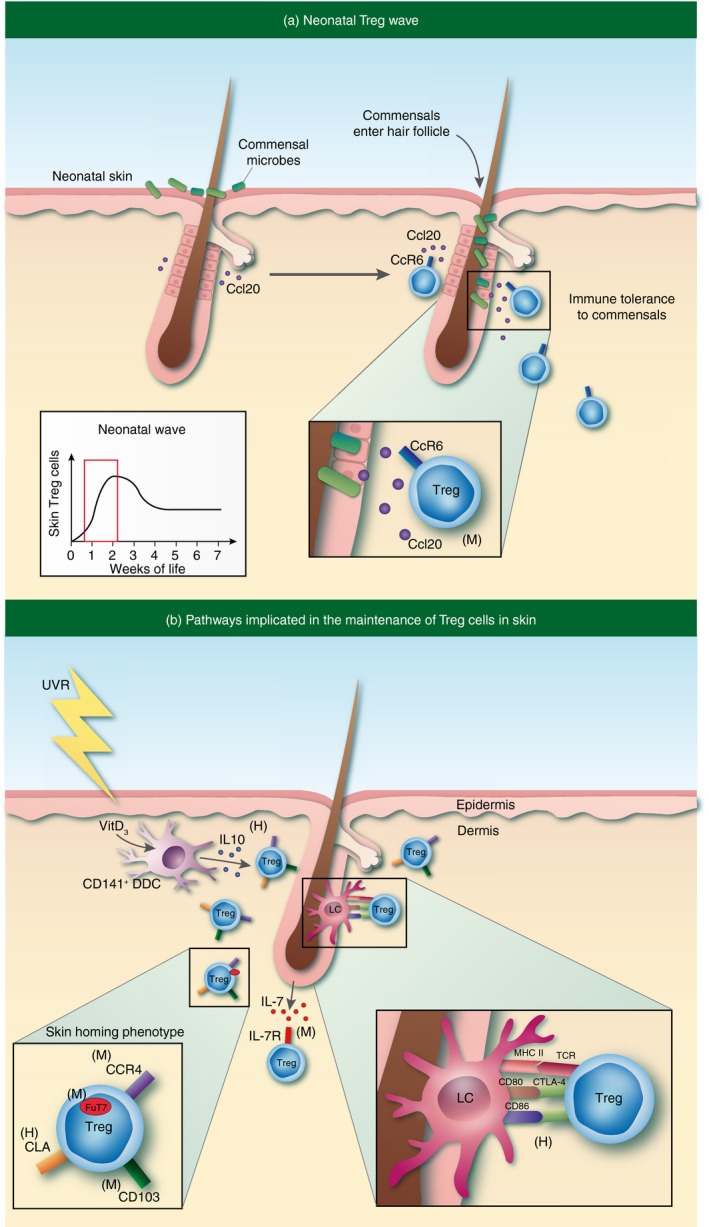
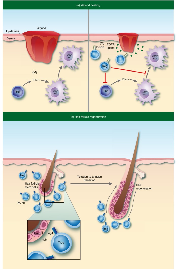
Foxp3+ CD4+ regulatory T (Treg) cells are a subset of immune cells that function to regulate tissue inflammation. Skin is one of the largest organs and is home to a large proportion of the body's Treg cells. However, relative to other tissues (such as the spleen and gastrointestinal tract) the function of Treg cells in skin is less well defined. Here, we review our understanding of how Treg cells migrate to skin and the cellular and molecular pathways required for their maintenance in this tissue. In addition, we outline what is known about the specialized functions of Treg cells in skin. Namely, the orchestration of stem cell-mediated hair follicle regeneration, augmentation of wound healing, and promoting adaptive immune tolerance to skin commensal microbes. A comprehensive understanding of the biology of skin Treg cells may lead to novel therapeutic approaches that preferentially target these cells to treat cutaneous autoimmunity, skin cancers and disorders of skin regeneration.
Keywords: autoimmunity; regulatory T cells; skin.
© 2017 John Wiley & Sons Ltd.
Figures
References
-
- Jordan MS, Boesteanu A, Reed AJ, Petrone AL, Holenbeck AE, Lerman MA et al Thymic selection of CD4+ CD25+ regulatory T cells induced by an agonist self‐peptide. Nat Immunol 2001; 2:301–6. - PubMed
-
- Sakaguchi S, Sakaguchi N, Asano M, Itoh M, Toda M. Immunologic self‐tolerance maintained by activated T cells expressing IL‐2 receptor α‐chains (CD25). Breakdown of a single mechanism of self‐tolerance causes various autoimmune diseases. J Immunol 1995; 155:1151–64. - PubMed
-
- Hori S, Nomura T, Sakaguchi S. Control of regulatory T cell development by the transcription factor Foxp3. Science 2003; 299:1057–61. - PubMed
-
- Fontenot JD, Gavin MA, Rudensky AY. Foxp3 programs the development and function of CD4+ CD25+ regulatory T cells. Nat Immunol 2003; 4:330–6. - PubMed
Publication types
MeSH terms
Substances
Grants and funding
LinkOut - more resources
Full Text Sources
Other Literature Sources
Medical
Research Materials
