Review
. 2017 Oct 20;27(12):823-838.
doi: 10.1089/ars.2017.7263. Epub 2017 Aug 10.

Redox Signaling in Diabetic Wound Healing Regulates Extracellular Matrix Deposition

Affiliations
Review

Redox Signaling in Diabetic Wound Healing Regulates Extracellular Matrix Deposition

Britta Kunkemoeller et al. Antioxid Redox Signal. .

Abstract

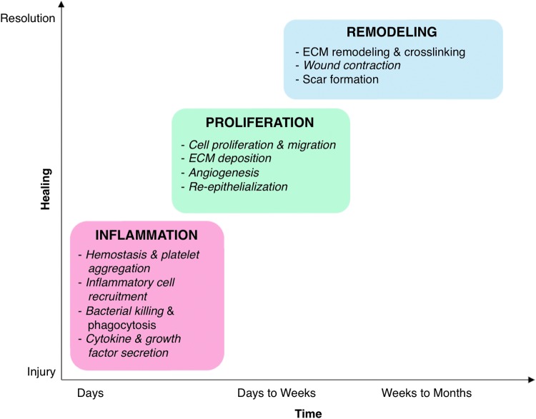
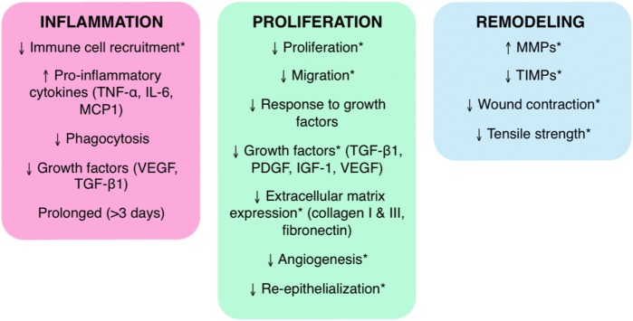
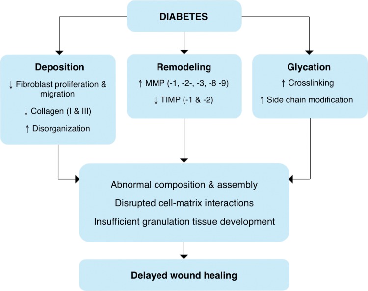
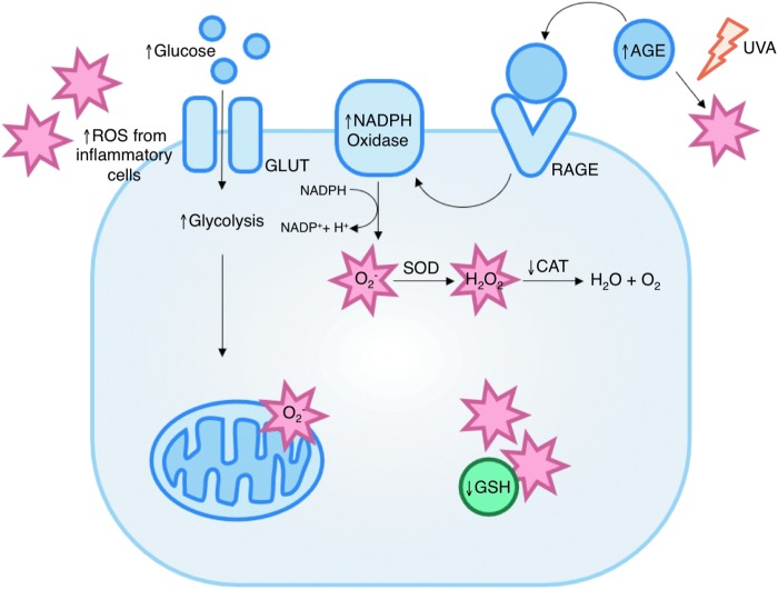
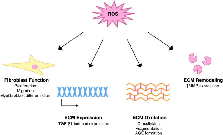
Significance: Impaired wound healing is a major complication of diabetes, and can lead to development of chronic foot ulcers in a significant number of patients. Despite the danger posed by poor healing, very few specific therapies exist, leaving patients at risk of hospitalization, amputation, and further decline in overall health. Recent Advances: Redox signaling is a key regulator of wound healing, especially through its influence on the extracellular matrix (ECM). Normal redox signaling is disrupted in diabetes leading to several pathological mechanisms that alter the balance between reactive oxygen species (ROS) generation and scavenging. Importantly, pathological oxidative stress can alter ECM structure and function.

Critical issues: There is limited understanding of the specific role of altered redox signaling in the diabetic wound, although there is evidence that ROS are involved in the underlying pathology.

Future directions: Preclinical studies of antioxidant-based therapies for diabetic wound healing have yielded promising results. Redox-based therapeutics constitute a novel approach for the treatment of wounds in diabetes patients that deserve further investigation. Antioxid. Redox Signal. 27, 823-838.

Keywords: collagen; diabetes; extracellular matrix; reactive oxygen species; wound healing.


Figures

<b>FIG. 1.</b>
FIG. 1.
Redox control of dermal wound healing. Normal wound healing occurs in three overlapping phases: inflammation, proliferation, and remodeling. Progression through these phases is highly regulated and coordinated by several mechanisms, including redox signaling. Both generation and scavenging of ROS, particularly H2O2, are critical to normal healing. The major processes regulated by redox signaling in each phase of healing are indicated in italics. ECM, extracellular matrix; H2O2, hydrogen peroxide; ROS, reactive oxygen species. To see this illustration in color, the reader is referred to the web version of this article at www.liebertpub.com/ars
<b>FIG. 2.</b>
FIG. 2.
Wound healing in diabetes. In contrast to normal healing, wound healing in diabetes is uncoordinated and spatiotemporally disorganized. Chronic diabetic wounds do not progress smoothly through inflammation, proliferation, and remodeling; they are instead characterized by an extended inflammation phase, a limited proliferation phase, and irregular remodeling. The critical changes in each phase of healing in diabetes are identified. Healing processes that involve ECM, a critical facilitator of healing because of its role as structural support and a mediator of cellular interactions, are indicated by asterisk (*). IGF-1, insulin-like growth factor-1; IL-6, interleukin-6; MCP-1, macrophage chemoattractant protein-1; MMP, matrix metalloproteinase; PDGF, platelet-derived growth factor; TGF-β1, transforming growth factor-β1; TIMP, tissue inhibitors of metalloproteinase; TNF-α, tumor necrosis factor-α; VEGF, vascular endothelial growth factor. To see this illustration in color, the reader is referred to the web version of this article at www.liebertpub.com/ars
<b>FIG. 3.</b>
FIG. 3.
ECM deposition is reduced in diabetes. Reduced deposition of ECM is characteristic of wound healing in diabetes. Masson's trichrome staining of mouse granulation tissue of healthy C57BL/6 mice (A) and diabetic db/db mice (B) reveals significantly reduced collagen deposition and maturation (blue). Wounds were explanted 14 days postinjury.
<b>FIG. 4.</b>
FIG. 4.
Changes in ECM in diabetes. The structure and function of the ECM are altered in diabetes via changes in fibroblast function, post-translational modification by glucose (glycation), and an imbalance of ECM deposition and remodeling. These changes influence matrix composition and assembly, cell–matrix interactions, and development of granulation tissue, and ultimately contribute to delayed wound healing in diabetes. Changes described in this figure have been found in human diabetic tissues and wounds. TIMP, tissue inhibitors of metalloproteinase. To see this illustration in color, the reader is referred to the web version of this article at www.liebertpub.com/ars
<b>FIG. 5.</b>
FIG. 5.
Sources of oxidative stress in diabetic wounds. Several mechanisms contribute to increased ROS levels in diabetes (indicated by stars). These include increased mitochondrial superoxide production, formation of advanced glycation end-products, increased activity of ROS-generating enzymes such as NADPH oxidase, and decreased expression of antioxidant enzymes and small molecules. AGE, advanced glycation end products; CAT, catalase; GLUT, glucose transporter; GSH, glutathione; RAGE, receptor for advanced glycation end products; SOD, superoxide dismutase. To see this illustration in color, the reader is referred to the web version of this article at www.liebertpub.com/ars
<b>FIG. 6.</b>
FIG. 6.
Excess mitochondrial superoxide production in diabetes. Hyperglycemia induces excess superoxide production by increasing the number of electron donors available to the electron transport chain. This increases the proton gradient past a critical level, and allows electron leakage (indicated by dashed lines) at complex I and CoQ. CoQ, coenzyme Q. To see this illustration in color, the reader is referred to the web version of this article at www.liebertpub.com/ars
<b>FIG. 7.</b>
FIG. 7.
Redox modulation of ECM. Redox signaling regulates ECM structure during normal wound healing, and excess ROS can cause pathological changes in ECM structure and function. TGF-β1, transforming growth factor-β1. To see this illustration in color, the reader is referred to the web version of this article at www.liebertpub.com/ars

References

    1. National Diabetes Statistics, 2011. Atlanta, GA: U.S. Department of Health and Human Services, Centers for Disease Control
    1. Global report on diabetes. Geneva, Switzerland: World Health Organization; 2014
    1. Acosta JB, del Barco DG, Vera DC, Savigne W, Lopez-Saura P, Guillen Nieto G, and Schultz GS. The pro-inflammatory environment in recalcitrant diabetic foot wounds. Int Wound J 5: 530–539, 2008 - PMC - PubMed
    1. Agren MS. and Werthen M. The extracellular matrix in wound healing: a closer look at therapeutics for chronic wounds. Int J Low Extrem Wounds 6: 82–97, 2007 - PubMed
    1. Aktunc E, Ozacmak VH, Ozacmak HS, Barut F, Buyukates M, Kandemir O, and Demircan N. N-acetyl cysteine promotes angiogenesis and clearance of free oxygen radicals, thus improving wound healing in an alloxan-induced diabetic mouse model of incisional wound. Clin Exp Dermatol 35: 902–909, 2010 - PubMed

MeSH terms