. 2017 Jul 20;547(7663):360-363.
doi: 10.1038/nature23010. Epub 2017 Jul 12.

Open and closed structures reveal allostery and pliability in the HIV-1 envelope spike

Affiliations

Open and closed structures reveal allostery and pliability in the HIV-1 envelope spike

Gabriel Ozorowski et al. Nature. .

Abstract

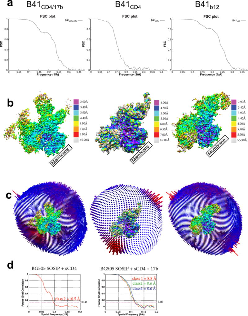
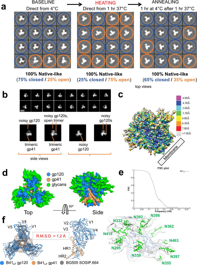
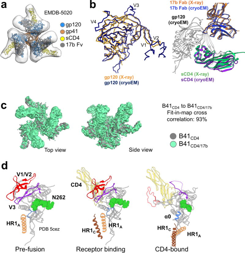
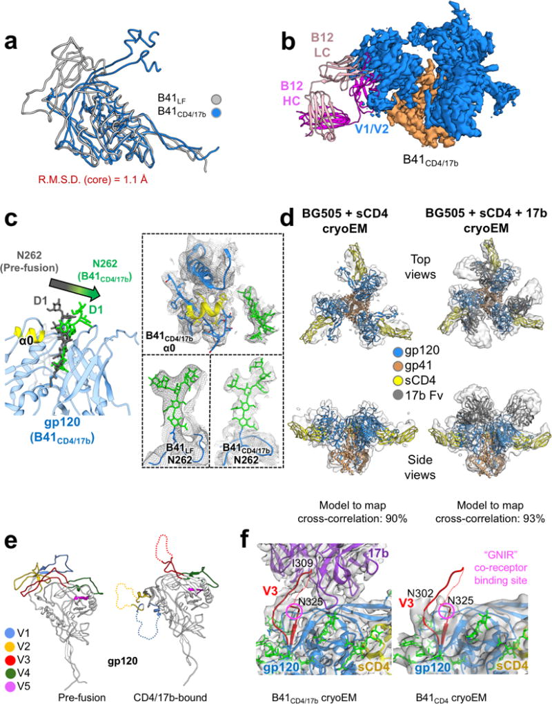
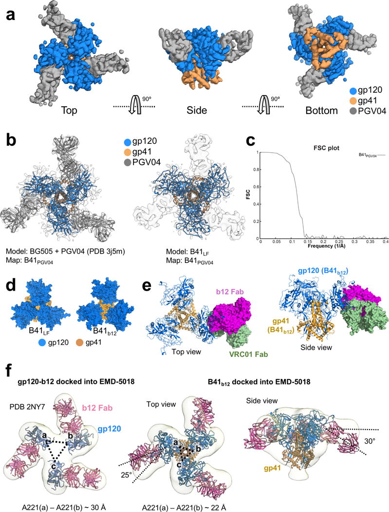
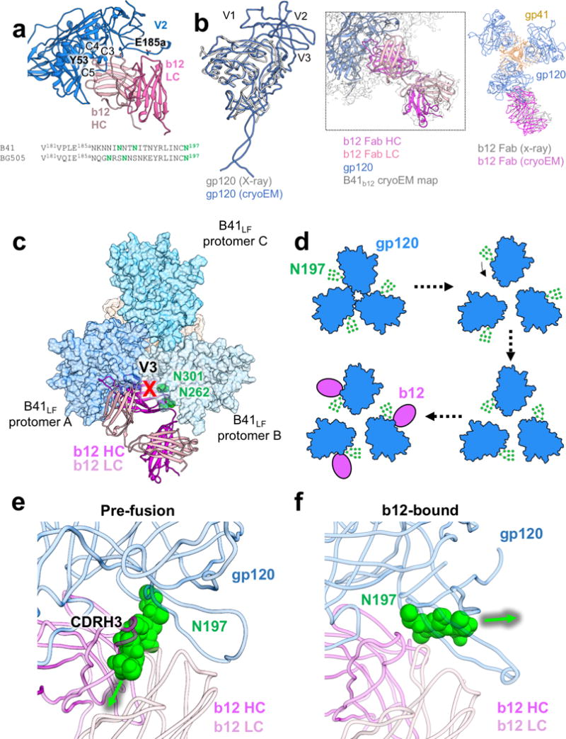
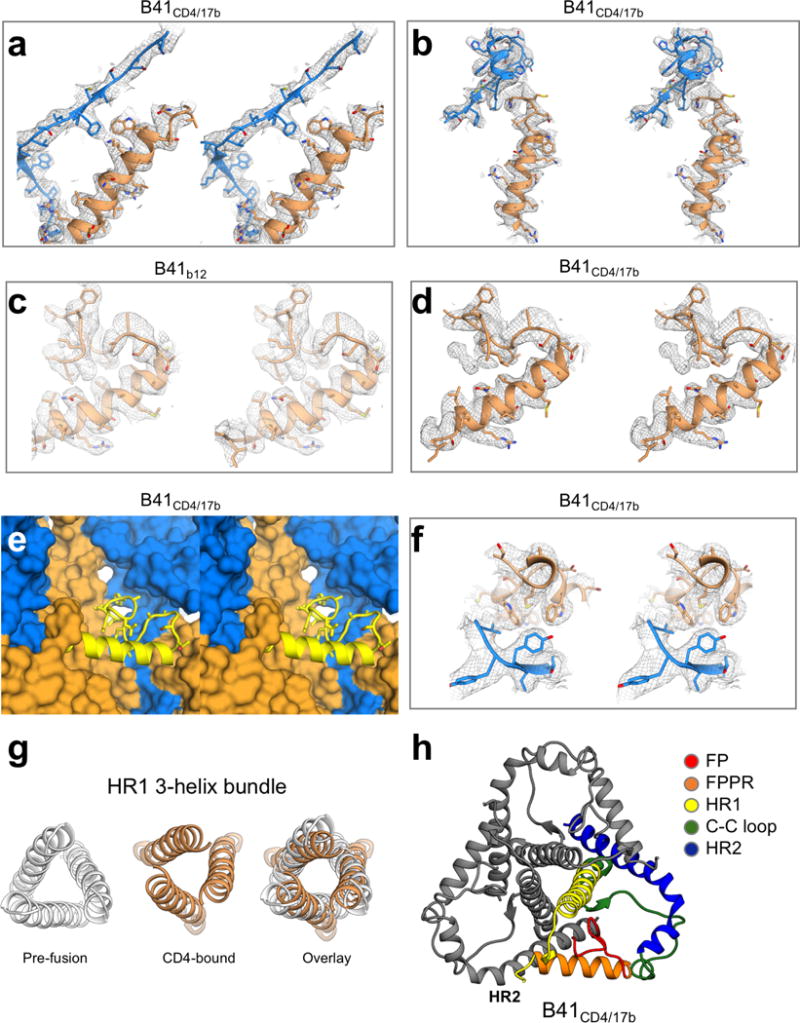
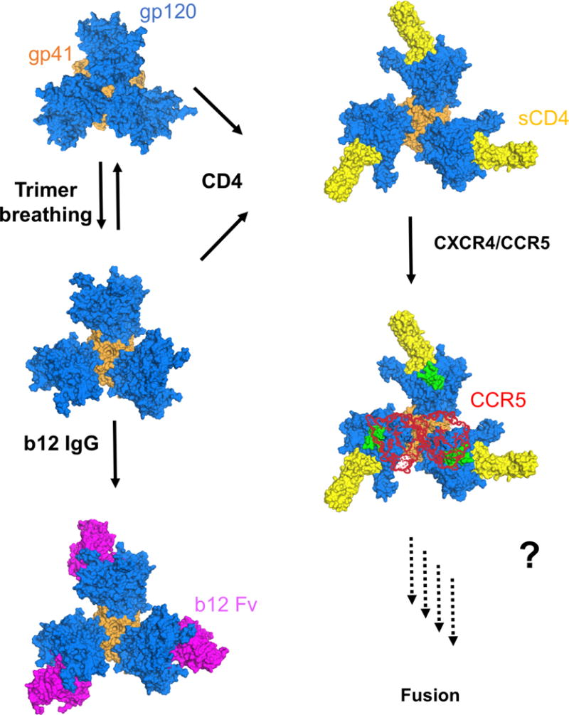
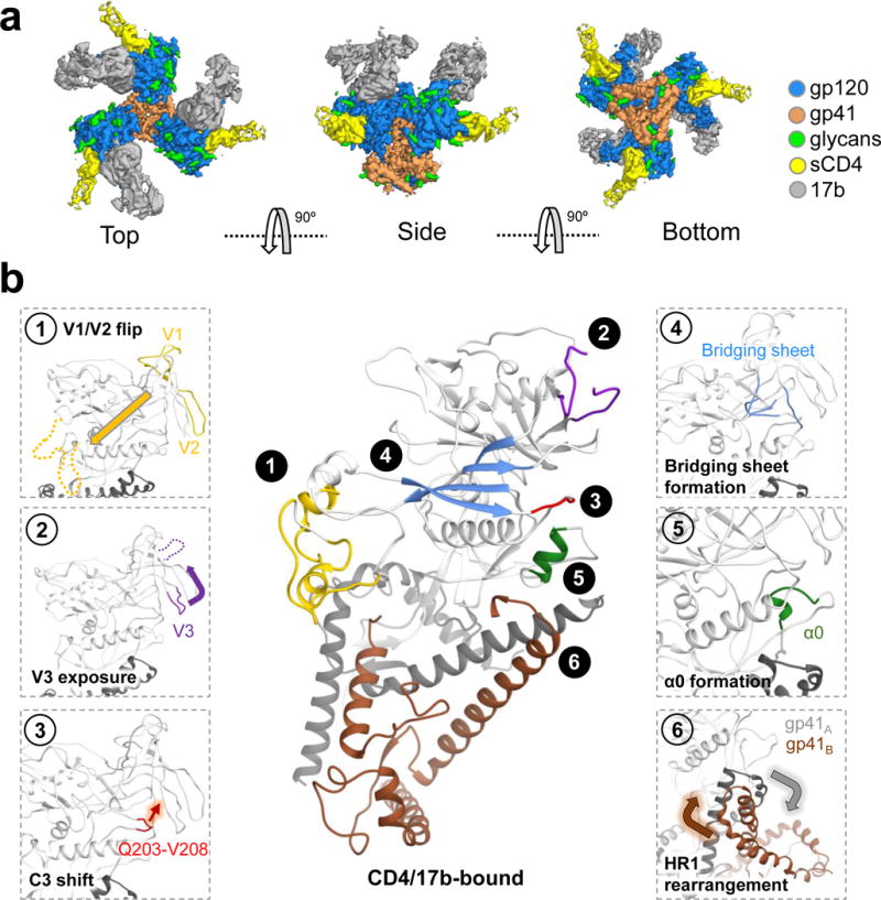
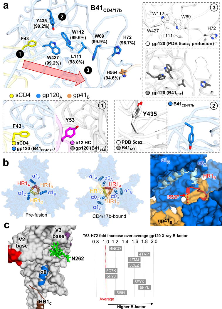
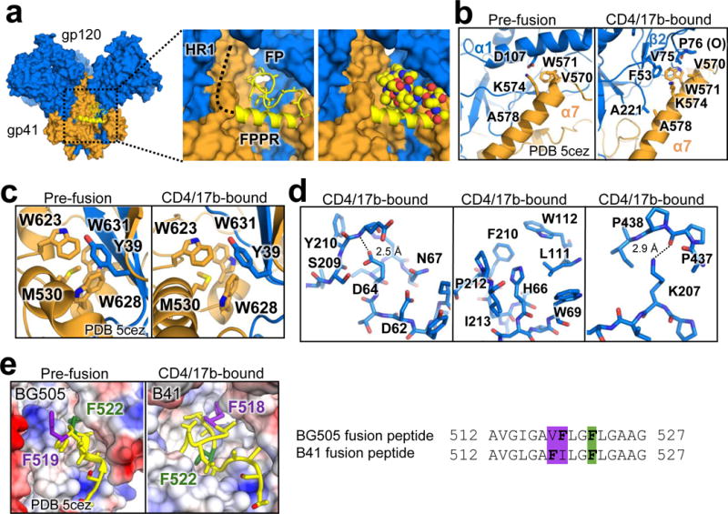
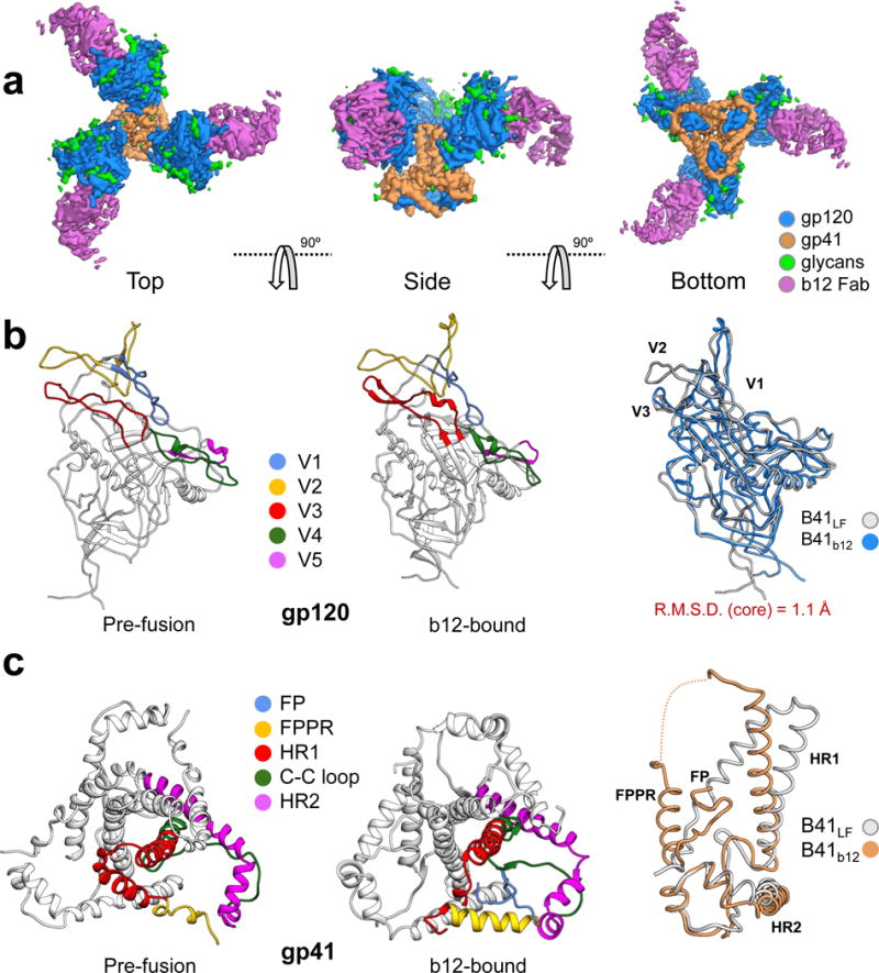
For many enveloped viruses, binding to a receptor(s) on a host cell acts as the first step in a series of events culminating in fusion with the host cell membrane and transfer of genetic material for replication. The envelope glycoprotein (Env) trimer on the surface of HIV is responsible for receptor binding and fusion. Although Env can tolerate a high degree of mutation in five variable regions (V1-V5), and also at N-linked glycosylation sites that contribute roughly half the mass of Env, the functional sites for recognition of receptor CD4 and co-receptor CXCR4/CCR5 are conserved and essential for viral fitness. Soluble SOSIP Env trimers are structural and antigenic mimics of the pre-fusion native, surface-presented Env, and are targets of broadly neutralizing antibodies. Thus, they are attractive immunogens for vaccine development. Here we present high-resolution cryo-electron microscopy structures of subtype B B41 SOSIP Env trimers in complex with CD4 and antibody 17b, or with antibody b12, at resolutions of 3.7 Å and 3.6 Å, respectively. We compare these to cryo-electron microscopy reconstructions of B41 SOSIP Env trimers with no ligand or in complex with either CD4 or the CD4-binding-site antibody PGV04 at 5.6 Å, 5.2 Å and 7.4 Å resolution, respectively. Consequently, we present the most complete description yet, to our knowledge, of the CD4-17b-induced intermediate and provide the molecular basis of the receptor-binding-induced conformational change required for HIV-1 entry into host cells. Both CD4 and b12 induce large, previously uncharacterized conformational rearrangements in the gp41 subunits, and the fusion peptide becomes buried in a newly formed pocket. These structures provide key details on the biological function of the type I viral fusion machine from HIV-1 as well as new templates for inhibitor design.


Conflict of interest statement

Competing financial interests

The authors declare no competing financial interests.

Figures

Extended Data Figure 1
Extended Data Figure 1. CryoEM statistics of B41CD4, B41CD4/17b and B41b12
(a) Fourier shell correlations. (b) Local resolution estimates. (c) Angular distribution plots. (d) Fourier shell correlations for BG505CD4 and BG505CD4/17b.
Extended Data Figure 2
Extended Data Figure 2. Cryo-EM reconstruction of ligand-free B41 SOSIP.664
(a) The “breathing” of B41 SOSIP.664 trimers is suggested by negative-stain 2D class averages with representative open (blue) and closed (orange) phenotypes highlighted. Sample temperature influences the percentage of open and closed phenotypes, and the open state is reversible. (b) CryoEM 2D class averages also suggest flexibility of the gp120 subunits relative to one another as evidenced by the blurring of gp120s in the class averages. (c) CryoEM reconstruction colored by local resolution for B41LF. (d) The segmented cryoEM map colored by component (gp120, gp41, glycans). The top view is defined as looking towards the viral membrane. (e) Fourier shell correlation (FSC) for B41LF. (f) Superposition of the x-ray structure of ligand-free BG505 SOSIP.664 (PDB 4zmj) onto a homology model of B41 SOSIP.664 refined into the cryoEM map (Cα R.M.S.D. ~1.2 Å). All three protomers are presented in the top view (left), while only a single protomer is shown in the side view for clarity (middle). Displayed is the V4 region and surrounding glycans (right).
Extended Data Figure 3
Extended Data Figure 3. Comparison of B41CD4/17b model
(a) Fitting of the cryoEM B41CD4/17b model into a cryoET reconstruction of surface-expressed Env in complex with sCD4 and 17b. (b) Alignment of gp120 in complex with CD4 and 17b from the cryoEM and x-ray crystallography models (PDB 1gc1). The Cα R.M.S.D. between our model and the x-ray structure of gp120 core in complex with sCD4 and 17b (PDB 1gc1) is ~1.5 Å, which is relatively low considering only 79% identity between the BG505 and B41 Env sequences. (c) Map fitting of B41CD4 into B41CD4/17b (low pass filtered to 5.2 Å) results in a cross correlation of 93%. (d) Cartoon representation of the major changes involved in the formation of the α0 helix. Pre-fusion model based on PDB 5CEZ.
Extended Data Figure 4
Extended Data Figure 4. Conformational differences between pre-fusion, b12-bound, and CD4-bound state
(a) Alignment of gp120 from the cryoEM models of B41LF and B41CD4/17b illustrates a significant displacement of the V1, V2, and V3 loops. (b) Fitting of the B41b12 model into the map of B41CD41/17b reveals a steric barrier created by b12 that prevents the translocation of V1/V2, which would clash with the antibody heavy chain. (c) Glycan N262 is repositioned away from the C1 domain of gp120 upon sCD4 binding as a result of V3 translocation (left). The relative positions of N262 in B41LF and B41CD4/17b are supported by continuous density (right). (d) Comparison of the cryoEM model of B41CD4/17b to cryoEM reconstructions of BG505CD4 and BG505CD4/17b. (e) Relative movements of variable loops between pre-fusion and CD4-bound states of B41 SOSIP (left). Loss of density for the V3 loop in both B41CD4 and B41CD4/17b maps suggests that V3 movement is in response to priming by CD4 and not caused by 17b or the co-receptor (right).
Extended Data Figure 5
Extended Data Figure 5. CryoEM reconstruction of B41 SOSIP.664 in complex with PGV04 Fab
(a) Segmented cryoEM map with components colored according to the key. (b) Docking of BG505 + PGV04 (PDB 3J5M) and B41LF models into the B41 + PGV04 reconstruction. Comparison with BG505 bound to PGV04 demonstrates a high degree of structural similarity (91% correlation between the two cryoEM maps), and docking of the B41LF model into the B41PGV04 map results in excellent agreement, with the B41LF backbone atoms falling into density as well as alignment of PNGS asparagine residues with glycan density (94% correlation between B41LF and B41PGV04 maps). (c) FSC of B41PGV04. (d) gp120 rotation and movement away from the central axis in the b12-bound state. (e) Comparison of VRC01 and b12 epitopes with respect to the open, b12-bound conformation of B41 SOSIP.664. (f) Docking of either the crystal structure of gp120-b12 or the B41b12 cryoEM model into the ECT reconstruction of HIV-1 BaL in complex with b12 (EMDB-5018) reveals differences in relative movement and rotation of gp120 from the trimer axis and ultimate position of b12.
Extended Data Figure 6
Extended Data Figure 6. Comparison of the B41b12 cryoEM model to the x-ray model of gp120 in complex with b12 Fab, and glycan repositionin
(a) Major contacts between b12 Fab and gp120 highlighted based on the cryoEM model. Sequence alignment between BG505 and B41 Env for a segment of V2, with N-linked glycans colored green. (b) Alignment of gp120 in complex with b12 from the cryoEM and x-ray crystallography models (PDB 2NYZ) (left). The crystal structure of b12 Fab docked into the B41b12 cryoEM map, revealing that the elbow angle is preserved (middle). When aligned to gp120, the x-ray model reveals a slightly different b12 angle of approach with respect to the cryoEM model (right). These differences may arise due to a more stable, neutralization resistant Tier-2 virus (B41) versus a more flexible lab-adapted Tier 1 BaL pseudovirus, the stabilizing SOSIP modifications used in the soluble constructs (although none are located at the CD4bs epitope or gp120 core), or the low resolution of the cryoET map. (c) The pre-fusion arrangement of gp120 does not allow for b12 binding to B41 SOSIP.664 due to clashes between the framework regions of the antibody and portions of a neighboring gp120 monomer (V3 and glycans). (d) Rotation of the N197 glycan requires the gp120 subunits to open up and move away from one another. (e,f) Glycan N197 in the b12 epitope acts as a steric barrier in the pre-fusion state and rearranges and moves away from the b12 epitope to allow for b12 binding.
Extended Data Figure 7
Extended Data Figure 7. CryoEM density of various stabilizing interactions in B41CD4/17b
(a, b) Stereo images of cryoEM density of specified contour levels for (a) K574-D107, and (b) the α0 HR1 cap of B41CD4/17b. (c, d) Fusion peptide and fusion peptide proximal region electron density for (c) B41b12 and (d) B41CD4/17b. (e) Stereo image of pocket protecting the fusion peptide in the CD4-bound state. (f) Stereo image of electron density of the Trp clasp region in B41CD4/17b. (g) HR1 3-helix bundle rearrangement between pre-fusion and CD4-bound states. (h) gp41 arrangement in B41CD4/17b. The FPPR and HR1N pack against regions of HR2 from two different protomers.
Extended Data Figure 8
Extended Data Figure 8. Overview of HIV-1 Env conformational states
Various biophysical data strongly suggest that the pre-fusion trimer is in an equilibrium of reversible open and closed states. b12 recognizes a more open state and traps the trimer in an irreversible intermediate state that can no longer play a role in host cell fusion. CD4, on the other hand, induces a stable, fusion intermediate that displays the co-receptor binding site and primes the fusion peptide to move to a more centralized location in the trimer interface. It is only after binding of CXCR4/CCR5 to CD4-bound Env that additional fusion steps occur, highlighted by the full formation of a three-helix bundle before final condensation into a six-helix bundle. When a dimer of CCR5 from a crystal structure is docked on top of the trimer structure, the N-termini of the two co-receptors are situated proximal to the co-receptor binding sites on gp120.
Figure 1
Figure 1. CryoEM reconstruction of B41 SOSIP.664 in complex with sCD4 and 17b Fab
(a) CryoEM map segmented by component. (b) CD4 binding results in a number of conformational changes in both the gp120 and gp41 regions of Env. Comparison of the CD4/17b-bound (middle) to pre-fusion states (side panels).
Figure 2
Figure 2. CD4/17b-bound B41 SOSIP.664 structural rearrangements that result in a stable fusion intermediate
(a) A network of hydrophobic and aromatic residues transmits the signal upon sCD4 binding that leads to the structural rearrangements in both gp120 and gp41. (b) The top of the HR1 helix is capped by the α1 helix of the same protomer in the pre-fusion state (left). CD4-binding results in the formation of helix α0, which now caps HR1 of a neighboring protomer (middle). A zoom in of the α0-HR1 interaction reveals that it is downstream to the engineered I559P mutation and likely a feature in native Env (right). (c) The D1 arm of glycan N262 rotates into a newly formed pocket on gp120 (left) positioned under the co-receptor binding site at the base of V3, providing a link between the co-receptor binding site and gp41. The α0 helix region is generally disordered in all published crystal structures of SOSIP trimers as evidenced by higher B-factors for this segment relative to the average gp120 value for each structure (right).
Figure 3
Figure 3. Additional stabilization resulting from CD4-bound rearrangements
(a) Space filling model of B41CD4/17b illustrating a newly created pocket that is now occupied by the fusion peptide. A close- up view of the pocket (center) and space-filling model (right). (b) K574 of gp41, which forms a salt bridge with D107 of gp120 in the pre-fusion state, now interacts with F53 of gp120. (c) The tryptophan clasp, previously described as a stabilizing feature of the pre-fusion state, is retained in the CD4-bound state. (d) Intra-protomer stabilizing interactions of helix α0 and β3-β4. (e) Two aromatic residues of the fusion peptide rearrange upon CD4-binding and fit in two pockets. Sequence alignment of the fusion peptides of BG505 (pre-fusion) and B41 (CD4-bound) with the aromatic residues highlighted. All pre-fusion model coordinates are based on PDB: 5CEZ.
Figure 4
Figure 4. CryoEM reconstruction and model of B41 SOSIP.664 in complex with b12 Fab
(a) CryoEM map segmented by component. (b) Comparison of gp120 variable loops before and after b12 binding; B41LF (left) and B41b12 (middle). Superposition of gp120 from pre-fusion and b12-bound states reveals only slight structural differences in the individual gp120 subunits (right). (c) Structural rearrangements in gp41 between the pre-fusion and b12-bound states include repacking of the HR1 three-helix bundle as well as ordering of the fusion peptide and fusion peptide proximal region (FPPR). Alignment onto HR2 of single gp41 chains from B41LF and B41b12 reveals a relative translocation of HR1 and rearrangement of the FP and FPPR (right).

Comment in

  • Viral infection: When two become one.
    York A. York A. Nat Rev Microbiol. 2017 Sep;15(9):511. doi: 10.1038/nrmicro.2017.85. Epub 2017 Jul 24. Nat Rev Microbiol. 2017. PMID: 28736446 No abstract available.

Similar articles

Cited by

References

    1. Harrison SC. Viral membrane fusion. Nat Struct Mol Biol. 2008;15:690–698. - PMC - PubMed
    1. Lindemann D, Steffen I, Pöhlmann S. In: Viral Entry into Host Cells. Pöhlmann Stefan, Simmons Graham., editors. Springer; New York: 2013. pp. 128–149.
    1. Lee JH, Ozorowski G, Ward AB. Cryo-EM structure of a native, fully glycosylated, cleaved HIV-1 envelope trimer. Science. 2016;351:1043. - PMC - PubMed
    1. Sanders RW, et al. A Next-Generation Cleaved, Soluble HIV-1 Env Trimer, BG505 SOSIP.664 gp140, Expresses Multiple Epitopes for Broadly Neutralizing but Not Non-Neutralizing Antibodies. PLoS Pathog. 2013;9:e1003618. - PMC - PubMed
    1. Burton DR, Hangartner L. Broadly Neutralizing Antibodies to HIV and Their Role in Vaccine Design. Annual Review of Immunology. 2016;34:635–659. - PMC - PubMed

Publication types

MeSH terms