. 2017 Jul 12;12(7):e0180032.
doi: 10.1371/journal.pone.0180032. eCollection 2017.

Ijuhya vitellina sp. nov., a novel source for chaetoglobosin A, is a destructive parasite of the cereal cyst nematode Heterodera filipjevi

Affiliations

Ijuhya vitellina sp. nov., a novel source for chaetoglobosin A, is a destructive parasite of the cereal cyst nematode Heterodera filipjevi

Samad Ashrafi et al. PLoS One. .

Abstract

Cyst nematodes are globally important pathogens in agriculture. Their sedentary lifestyle and long-term association with the roots of host plants render cyst nematodes especially good targets for attack by parasitic fungi. In this context fungi were specifically isolated from nematode eggs of the cereal cyst nematode Heterodera filipjevi. Here, Ijuhya vitellina (Ascomycota, Hypocreales, Bionectriaceae), encountered in wheat fields in Turkey, is newly described on the basis of phylogenetic analyses, morphological characters and life-style related inferences. The species destructively parasitises eggs inside cysts of H. filipjevi. The parasitism was reproduced in in vitro studies. Infected eggs were found to harbour microsclerotia produced by I. vitellina that resemble long-term survival structures also known from other ascomycetes. Microsclerotia were also formed by this species in pure cultures obtained from both, solitarily isolated infected eggs obtained from fields and artificially infected eggs. Hyphae penetrating the eggshell colonised the interior of eggs and became transformed into multicellular, chlamydospore-like structures that developed into microsclerotia. When isolated on artificial media, microsclerotia germinated to produce multiple emerging hyphae. The specific nature of morphological structures produced by I. vitellina inside nematode eggs is interpreted as a unique mode of interaction allowing long-term survival of the fungus inside nematode cysts that are known to survive periods of drought or other harsh environmental conditions. Generic classification of the new species is based on molecular phylogenetic inferences using five different gene regions. I. vitellina is the only species of the genus known to parasitise nematodes and produce microsclerotia. Metabolomic analyses revealed that within the Ijuhya species studied here, only I. vitellina produces chaetoglobosin A and its derivate 19-O-acetylchaetoglobosin A. Nematicidal and nematode-inhibiting activities of these compounds have been demonstrated suggesting that the production of these compounds may represent an adaptation to nematode parasitism.


Conflict of interest statement

Competing Interests: The authors have declared that no competing interests exist.

Figures

Fig 1
Fig 1. Cysts and eggs of Heterodera filipjevi naturally infected with Ijuhya vitellina, and pure cultures obtained from the infected eggs.
(A) Symptomatic, reddish dotted nematode cysts. (B, C) Nematode eggs accommodating reddish microsclerotia. (D, E) Microsclerotial tissue developing inside juveniles. (F) A six-month-old culture that developed from a single infected nematode egg. (G) Surface of colony showing reddish microsclerotia arranged in concentric rings. (H, I) Two-month-old cultures on PDA and CMA. Scale bars: A = 0.5 mm, B = 30 μm, C-E = 50 μm, F = 1 cm (also applying for H, I), G = 400 μm.
Fig 2
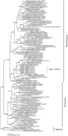
Fig 2. Bayesian inference of phylogenetic relationships of selected taxa of the Bionectriaceae and Nectriaceae (Hypocreales) based on LSU sequences.
Numbers above nodes are estimates of a posteriori probabilities (≥ 0.9) / NJB and MLB values (≥70%). The topology was rooted with Aschersonia placenta, Balansia henningsiana, B. pilulaeformis, and Hypocrella nectrioides, (Hypocreales).
Fig 3
Fig 3. Bayesian inference of infrageneric phylogenetic relationships within Ijuhya based on act, ITS, LSU, rpb1, and ß-tub sequences.
Numbers above nodes are estimates of a posteriori probabilities (≥ 0.9) / NJB and MLB (≥ 70%). The topology was rooted with three distantly related ‘Ijuhya’ species (‘Ijuhyaantillana, I. dentifera, and ‘Ijuhyaoenanthicola).
Fig 4
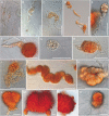
Fig 4. Light micrographs of Ijuhya vitellina, formation of microsclerotia.
(A-F) Transformation of hyphae into (A-D) chlamydospore or dictyochlamydospore-like structures, and (E, F) microsclerotia. (G-I) Coiling or coalescence of dictyochlamydospore-like structures. (J) Microsclerotia densely arranged in a chain. (K-N) Pigmentation first observed (K) in cell walls, and later (L-M) intensifying throughout microsclerotia. (O) A single microsclerotium inoculated on agar surface developing hyphae. A-I, K-N: from PDA, J: from CMA, O: from PDA 1/3. Scale bars: (A, C, E-I, K, L, O) = 30 μm; B, J = 50 μm; D = 200 μm; (M, N) = 10 μm.
Fig 5
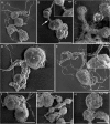
Fig 5. SEM micrographs of microsclerotia formed by Ijuhya vitellina.
(A) Filamentous hyphae developing into multicellular structures. (B) Intercalary formed dictyochlamydospores connected by hyphae (arrowed). (C) Detail of intercalary multicellular structures of microsclerotia. (D, E) Hyphae transformed into chlamydospore-like structures and microsclerotia. (F) Terminally formed microsclerotium. (G) Moniliform arrangement of microsclerotia. (H) Detail of microsclerotia illustrating a multicellular surface that forms a textura angularis. Scale bars: A = 100 μm; (B, E-H) = 50 μm; (C, D) = 30 μm.
Fig 6
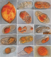
Fig 6. Light micrographs of the infection and colonisation process of Ijuhya vitellina in cysts and eggs of Heterodera filipjevi.
(A) Symptomatic cyst, reddish-dotted due to eggs containing reddish, globose microsclerotia. (B-E) Early colonisation of nematode eggs by hyphae becoming chlamydospore- and dictychlamydospore-like to develop microsclerotia inside eggs. (F, G) Dictyochlamydospore-like structures and small microsclerotia. (H) Hyphae penetrating through the eggshell by forming appressorium-like structure (arrows). (I-K) Development of the fungus inside nematodes eggs: (I) Formation of thick-walled hyphal cells, later (J-K) transforming into microsclerotia. The arrow in (J) points at the nematode stylet; in (K) at immature microsclerotium. (L) Egg with mature microsclerotium. (M-N) Near-identical cells of microsclerotium formed in (M) egg and (N) pure culture, forming a textura angulari in optical sections. Material obtained from (B-G, M) infected cysts directly placed and incubated on fungal colony, (H-L) slide cultures, (N) OA. Scale bars: A = 300 μm; (B-N) = 30 μm.
Fig 7
Fig 7
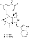
Structures of chaetoglobosin A (1) and 19-O-acetylchaetoglobosin A (2).

References

    1. Kühn J. Vorläufiger Bericht über die bisherigen Ergebnisse der seit dem Jahre 1875 im Auftrage des Vereins für Rübenzucker-Industrie ausgeführten Versuche zur Ermittlung der Ursache der Rübenmüdigkeit des Bodens und zur Erforschung der Natur der Nematoden. Z Ver Rübenzucker-Ind. 1877;27:452–7.
    1. Stirling GR. Biological control of plant-parasitic nematodes: soil ecosystem management in sustainable agriculture Second Edition ed. Stirling GR, editor. Wallingford, UK: CABI; 2014. xxiv + 510 p.
    1. Goffart H. Untersuchungen am Hafernematoden Heterodera schachtii Schm. unter besonderer Brücksichtigung der schleswig-holsteinischen Verhältnisse I. Arbeiten aus der Biologischen Reichsanstalt für Land- und forstwirtschaft Berlin-Dahlem. 1932;20:1–28.
    1. Kerry BR. A fungus associated with young females of the cereal cyst-nematode, Heterodera avenae. Nematologica. 1974;20(2):259–61. doi: 10.1163/187529274x00230 - DOI
    1. Kerry BR, Crump DH. Two fungi parasitic on females of cystnematodes (Heterodera spp.). Transactions of the British Mycological Society. 1980;74(1):119–25.

LinkOut - more resources