. 2017 Jul 12;12(7):e0180225.
doi: 10.1371/journal.pone.0180225. eCollection 2017.

BRG1 promotes hepatocarcinogenesis by regulating proliferation and invasiveness

Affiliations

BRG1 promotes hepatocarcinogenesis by regulating proliferation and invasiveness

Benedikt Kaufmann et al. PLoS One. .

Abstract

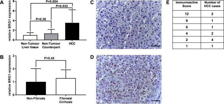
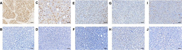
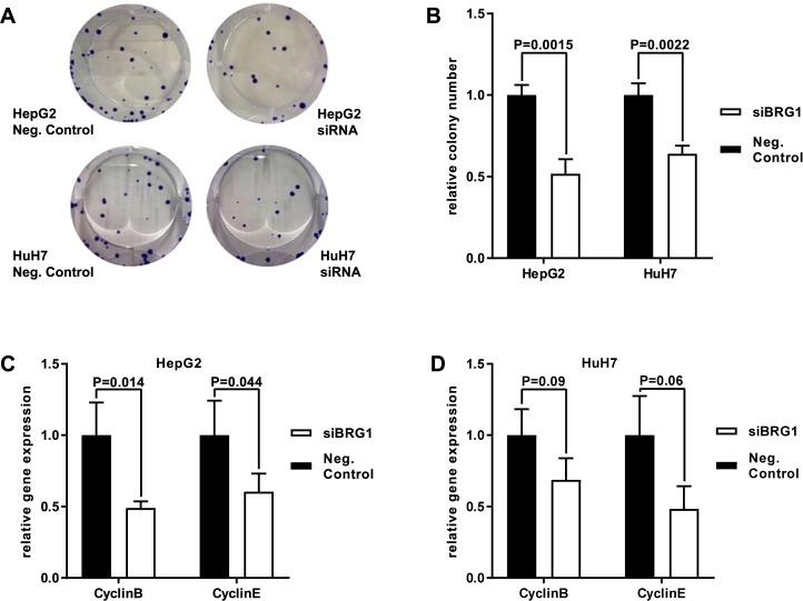
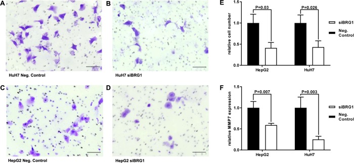
The chromatin remodeler complex SWI/SNF plays an important role in physiological and pathological processes. Brahma related gene 1(BRG1), a catalytic subunit of the SWI/SNF complex, is known to be mutated in hepatocellular carcinoma (HCC). However, its role in HCC remains unclear. Here, we investigate the role of BRG1 on cell growth and invasiveness as well as its effect on the expression of putative target genes. Expression of BRG1 was examined in human liver tissue samples and in HCC cell lines. In addition, BRG1 was silenced in human HCC cell lines to analyse cell growth and invasiveness by growth curves, colony formation assay, invasion assay and the expression of putative target genes. BRG1 was found to be significantly increased in HCC samples compared to non-HCC samples. In addition, a declined proliferation rate of BRG1-silenced human HCC cell lines was associated with a decrease of expression of cyclin family members. In line with a decreased invasiveness of BRG1-siRNA-treated human HCC cell lines, down-regulation of MMP7 was detected. These results support the hypothesis that overexpression of BRG1 increases cell growth and invasiveness in HCC. Furthermore, the data highlight cyclin B, E and MMP7 to be associated with BRG1 during hepatocarcinogenesis.


Conflict of interest statement

Competing Interests: The authors have declared that no competing interests exist.

Figures

Fig 1
Fig 1. Analysis of BRG1 expression in HCC by qRT-PCR and immunohistochemical staining.
(A,B) BRG1 is overexpressed significantly in HCC tissue (n = 13) compared to non-tumour counterpart (n = 10) respectively non-tumour liver tissue of patients not suffering from HCC (n = 13). A more detailed analysis of non-tumour liver tissue revealed no difference of BRG1 expression between tissue of non-fibrosis (n = 13) and fibrosis/cirrhosis (n = 10). Shown are the relative expression levels, non-tumour liver tissue respectively non-fibrosis tissue were standardised as 1. (C) Normal hepatocytes are showing no expression of BRG1. (D) Positive BRG1 staining in HCC tissue. (G) Analysis of BRG1 expression in immunohistochemistry by immunoreactive score. A varying degree of BRG1 expression in HCC was found ranging from a minor score of 1 to a maximum score of 12. The determined scores were evenly distributed. Differences were due to a high variety of intensity as well as the percentage of positive stained cells. Scale bar represents 50μm.
Fig 2
Fig 2. Immunohistochemical staining of BRG1 in HCC.
(A-J) BRG1 is expressed in HCC tissue (A,C,E,G,I) but not in non-tumour tissue counterpart (B,D,F,H,J). (A,C,E,G,I) HCC tissue is showing different expression levels of BRG1 due to a high variety of intensity as well as the percentage of positive stained cells. Scale bar represents 50μm.
Fig 3
Fig 3. BRG1 knockdown impairs proliferation.
(A,B) Human HCC cell lines HepG2 and HuH7 showed expression of BRG1 on mRNA and protein level. (A) After transfection, a highly significant down-regulation of mRNA levels of BRG1 was achieved for both HepG2 (P<0.001) and HuH7 (P<0.001) cell lines, n = 4. (B) Analysed by Western Blot, protein expression was also decreased in HepG2 and HuH7 cell lines after transfection, n = 3. Proliferation was analysed by growth curves for both human HCC cell lines HepG2 and HuH7. (C,D) Growth curves, n = 3 and (E,F) BRG1 expression at different time points after transfection, n = 3. Growth curves of HepG2 (C) and HuH7 (D) cells revealed a significant decrease of proliferation as long as BRG1 is suppressed significantly (E,F). 10 days after transfection proliferation rates began to equalize (C,D) and siRNA targeting BRG1 started to lose its impact (E,F). (E,F) Negative control was standardised as 1.
Fig 4
Fig 4. BRG1 knockdown impairs colony formation and modulates cyclin family.
(A,B) Colony formation assay was performed to analyse proliferation of both human HCC cell lines HepG2 and HuH7. Colony formation was reduced significantly in both cell lines, HepG2 and HuH7, n = 3. (C) Analysis of mRNA expression by qRT-PCR showed a significant down-regulation of cyclinB and cyclinE expression in BRG1-suppressed HepG2 cells at the time point of 40h after transfection, n = 4. (D) HuH7 cells showed a trend towards lower expression though this was not significant, n = 4. Negative control was standardised as 1.
Fig 5
Fig 5. BRG1 knockdown impairs invasiveness and modulates MMP7.
Invasion assay was performed to analyse the role of BRG1 for invasive ability of human HCC cell lines. (A-E) In both cell lines down-regulated BRG1 expression impaired invasive ability. (F) Analysis of mRNA levels after down-regulation of BRG1 showed a significant decrease of MMP7 expression for both cell lines 40h after transfection. Negative control was standardised as 1. Scale bar represents 100μm.

References

    1. Jemal A., Bray F., Center M.M., Ferlay J., Ward E., Forman D. Global cancer statistics. CA Cancer J Clin. 2011; 61: 69–90. doi: 10.3322/caac.20107 - DOI - PubMed
    1. El-Serag H. B., Rudolph K. L. Hepatocellular carcinoma: epidemiology and molecular carcinogenesis. Gastroenterology. 2007; 132: 2557–2576. doi: 10.1053/j.gastro.2007.04.061 - DOI - PubMed
    1. Lee J.-S. The mutational landscape of hepatocellular carcinoma. Clin Mol Hepatol. 2015; 21: 220–229. doi: 10.3350/cmh.2015.21.3.220 - DOI - PMC - PubMed
    1. Totoki Y., Tatsuno K., Covington K.R., Ueda H., Creighton C.J., Kato M., et al. Trans-ancestry mutational landscape of hepatocellular carcinoma genomes. Nat Genet. 2014; 46: 1267–1273. doi: 10.1038/ng.3126 - DOI - PubMed
    1. Kadoch C., Hargreaves D. C., Hodges C., Elias L., Ho L., Ranish J., et al. Proteomic and bioinformatic analysis of mammalian SWI/SNF complexes identifies extensive roles in human malignancy. Nat Genet. 2013; 45: 592–601. doi: 10.1038/ng.2628 - DOI - PMC - PubMed

MeSH terms