. 2017 Jul 11;12(7):e0181199.
doi: 10.1371/journal.pone.0181199. eCollection 2017.

Gallic acid/hydroxypropyl-β-cyclodextrin complex: Improving solubility for application on in vitro/ in vivo Candida albicans biofilms

Affiliations

Gallic acid/hydroxypropyl-β-cyclodextrin complex: Improving solubility for application on in vitro/ in vivo Candida albicans biofilms

Guilherme Rodrigues Teodoro et al. PLoS One. .

Abstract

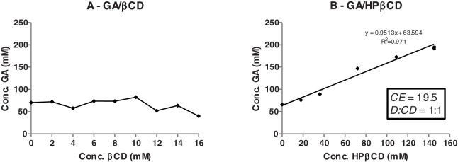
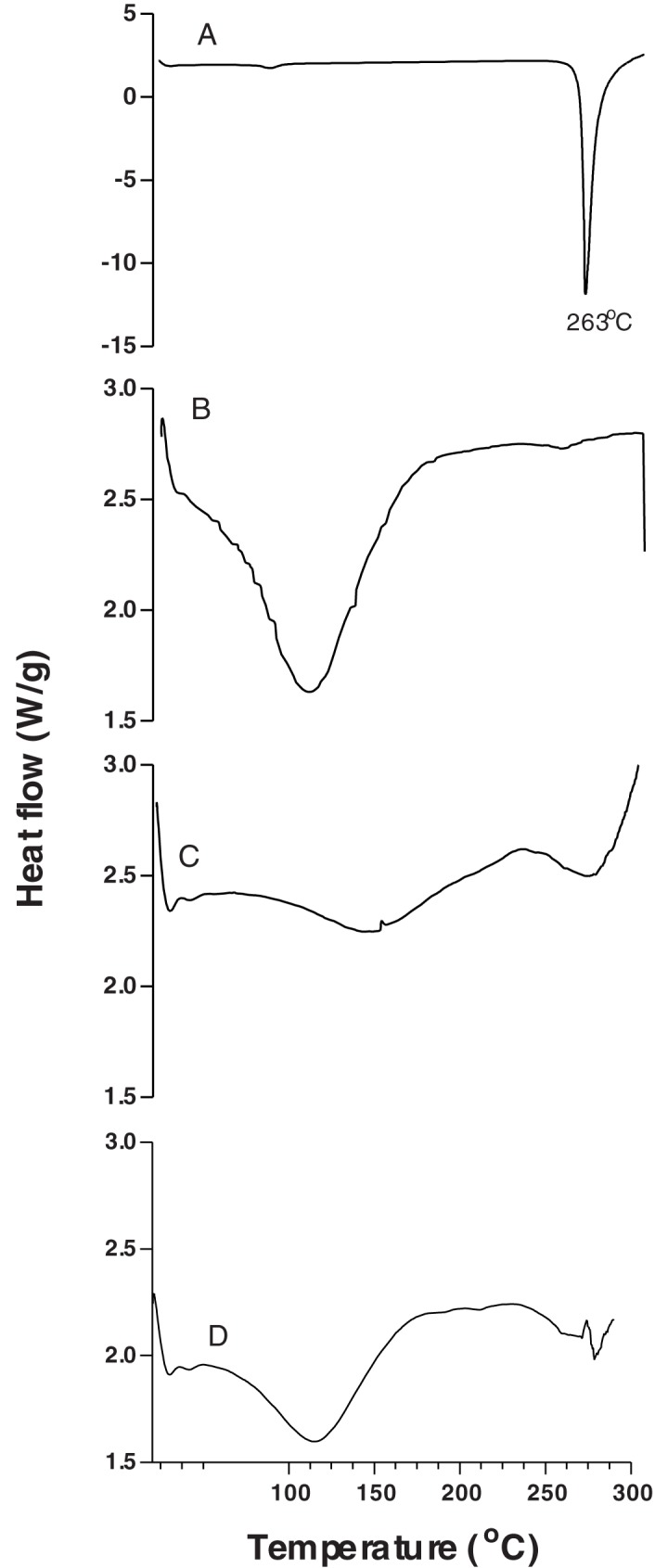
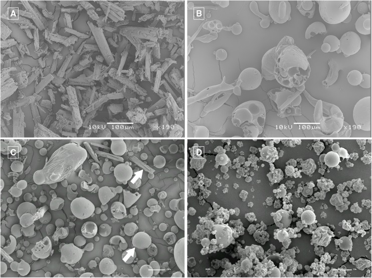
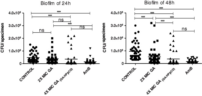
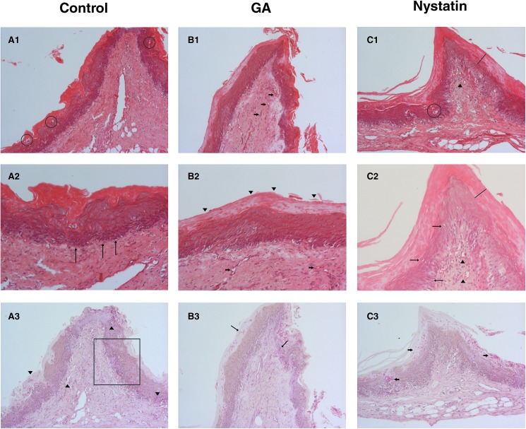
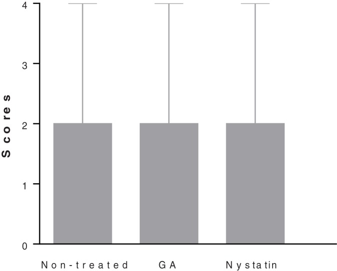
The aim of this study was to increase the solubility of gallic acid (GA) for the treatment of Candida albicans biofilm, which is very difficult to treat and requires high drug concentrations. Cyclodextrins (CDs) were used for this purpose. Complexes were evaluated by phase-solubility studies, prepared by spray drying and characterized by drug loading, scanning electron microscopy (SEM) and differential scanning calorimetry (DSC). The complexes were tested on C. albicans biofilm using in vitro and in vivo models. HPβCD formed soluble inclusion complexes with GA. The percentage of GA in GA/HPβCD was 10.8 ± 0.01%. The SEM and DSC analyses confirmed the formation of inclusion complexes. GA/HPβCD maintained the antimicrobial activity of the pure GA. GA/HPβCD was effective on C. albicans biofilms of 24 and 48h. The in vivo results showed an anti-inflammatory activity of GA/HPβCD with no difference in invading hypha counting among the groups. This study encourages the development of new antifungal agents.


Conflict of interest statement

Competing Interests: The authors have declared that no competing interests exist.

Figures

Fig 1
Fig 1. Phase solubility diagrams.
Gallic acid (GA) in aqueous solutions of βCD (A) and HPβCD (B). CE is the complexation efficiency and D:CD is the molar ratio between GA and HPβCD.
Fig 2
Fig 2. DSC thermograms.
Gallic acid (GA) raw material (A), HPβCD (B), physical mixture between GA and HPβCD (C) and GA/HPβCD spray-dried particle (D).
Fig 3
Fig 3. SEM micrographs.
Gallic acid (GA) 190X magnification (A), HPβCD 190X magnification (B), physical mixture of GA and HPβCD 150X magnification (C) and GA/HPβCD spray-dried microparticles, 2,000X magnification (D). In Fig C the upper arrow points out to the GA morphology and the lower arrow points out to the HPβCD one.
Fig 4
Fig 4
Effect of galic acid (GA) on C. albicans biofilm of 24 h (A) and 48h (B).Biofilms were exposed to 2 and 4 times the MIC (10 and 20 mg/mL) of GA for 5 min or to amphotericin B (AmB, 2 μg ml-1). Four times the MIC of GA (20 mg/mL) was insoluble in water and GA/HPβCD spray-dried microparticles were tested. Control refers to negative control. Each point represents the mean of the data from three independent experiments (n = 45); bars represent the standard deviation (SD). Kruskal-Wallis test and Dunn's Multiple Comparison post hoc test were used to compare the treatment groups against the negative control.***, P ≤ 0.001; ns, not significant.
Fig 5
Fig 5. Antifungal activity on oral candidiasis in rats.
Control Group (A), GA/HPβCD spray-dried microparticles group (B) and Nystatin group (C). (A1) Histologic sections stained by Hematoxylin and eosin (HE) method, 100X magnification, exhibiting hyperkeratosis, and micro-abscesses formation (circle) in the epithelium that is also acanthotic. The fibrous connective tissue has wide vessels surrounded by a moderate inflammatory infiltrate. (A2) PAS staining, 100X magnification, where C. albicans yeast and mostly hyphae invade the epithelium reaching the connective tissue (arrowheads). (A3) HE staining, 200X magnification. Inflammatory epithelial alterations as hyperkeratosis, epithelial stratification loss (square), and basal layer disorganization (arrows) are evident. (B1) Histologic sections stained by hematoxylin and eosin (HE) method, 100X magnification, exhibiting hyperkeratosis, and acanthosis of the epithelium that covers a hyper-vascularized (arrows) connective tissue. (B2) HE staining, 200X magnification. Reactive hyperkeratosis of the epithelium (arrowheads) was the only remarkable inflammatory induced alteration for the GA/HPβCD group. Some congest vessels can be seen in the connective tissue (arrows). (B3) PAS staining, 100X magnification, where C. albicans yeast and mostly hyphae invade the epithelium, some of them reaching the connective tissue (arrows). (C1) Histologic sections stained by hematoxylin and eosin (HE) method, 100X magnification, hyperkeratosis (line), micro-abscess formation (circle), and acanthosis of the epithelium that covers a hyper-vascularized (arrowheads) connective tissue can be seen. (C2) PAS staining, 100X magnification where large groups of Candida albicans yeast and hyphae can be noted. Though most of them are limited to the keratin layer, some invade the epithelium (arrows). (C3) HE staining, 200X magnification, where epithelial hyperkeratosis (line) is evident. We can see poor inflammatory alteration (arrows), as hydropic degeneration and duplication of the basal layer. Some congest vessels can be seen in the connective tissue (arrowheads) surrounded by moderate inflammatory infiltrate.
Fig 6
Fig 6. Analysis of hypha counting.
Median with range of the attributed scores for each group after hypha counting.

Similar articles

Cited by

References

    1. Williams DW, Jordan RP, Wei XQ, Alves CT, Wise MP, Wilson MJ, et al. Interactions of with host epithelial surfaces. J Oral Microbiol. 2013;5: 22434. - PMC - PubMed
    1. Pfaller MA, Diekema DJ. Epidemiology of invasive candidiasis: a persistent public health problem. Clinical microbiology reviews. 2007;20(1):133–63. doi: 10.1128/CMR.00029-06 - DOI - PMC - PubMed
    1. Uppuluri P, Pierce CG, López-Ribot JL. Candida albicans biofilm formation and its clinical consequences. Future Microbiology. 2009;4(10):1235–7. doi: 10.2217/fmb.09.85 - DOI - PMC - PubMed
    1. Douglas LJ. Candida biofilms and their role in infection. Trends in Microbiology. 2003;11(1):30–6. doi: https://doi.org/10.1016/S0966-842X(02)00002-1 - DOI - PubMed
    1. Bujdáková H. Management of Candida biofilms: state of knowledge and new options for prevention and eradication. Future Microbiology. 2016;11(2):235–51. doi: 10.2217/fmb.15.139 - DOI - PubMed

MeSH terms

LinkOut - more resources