. 2017 Jul 11;20(2):439-450.
doi: 10.1016/j.celrep.2017.06.041.

Viral Activation of Heparanase Drives Pathogenesis of Herpes Simplex Virus-1

Affiliations

Viral Activation of Heparanase Drives Pathogenesis of Herpes Simplex Virus-1

Alex M Agelidis et al. Cell Rep. .

Abstract

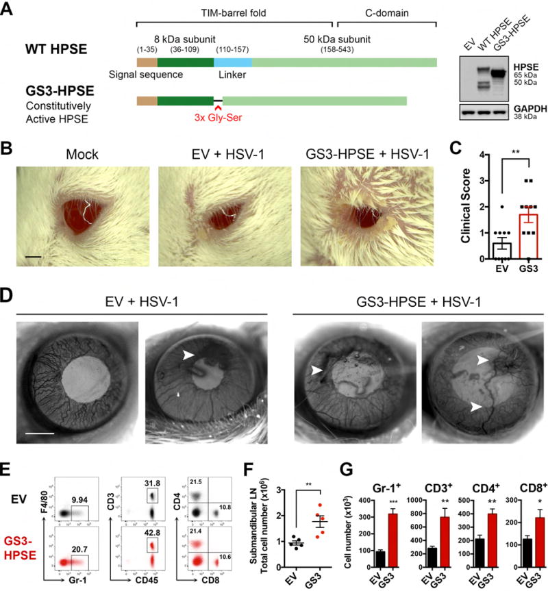
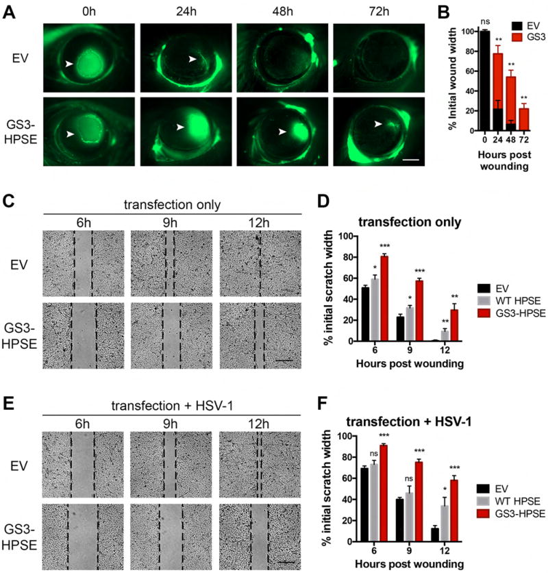
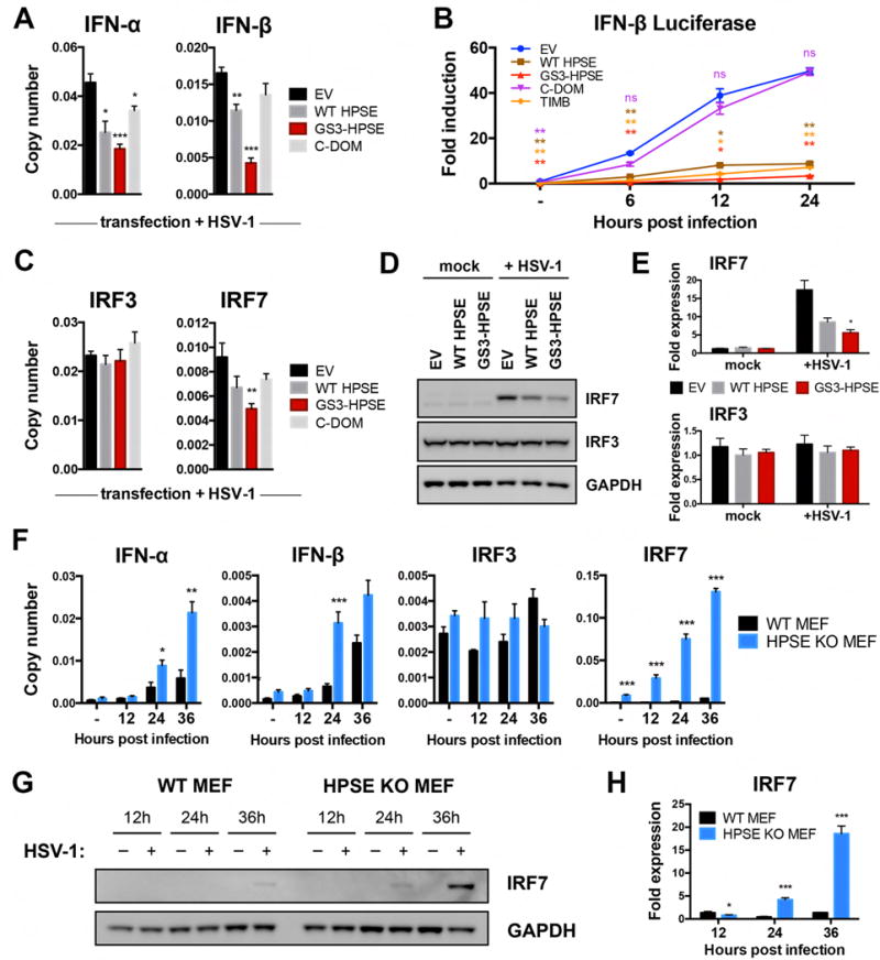
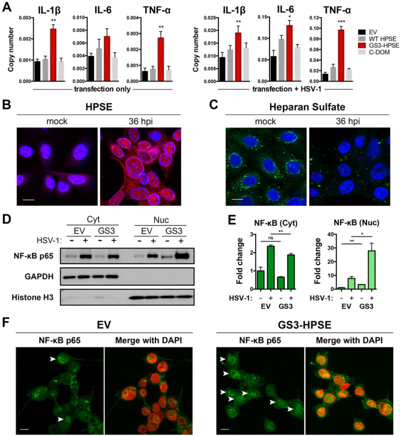
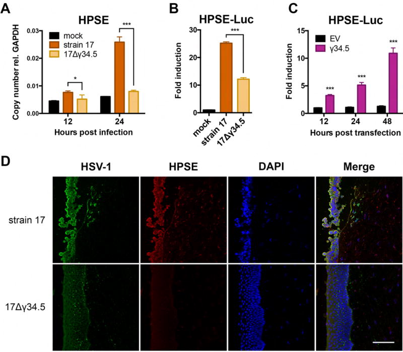
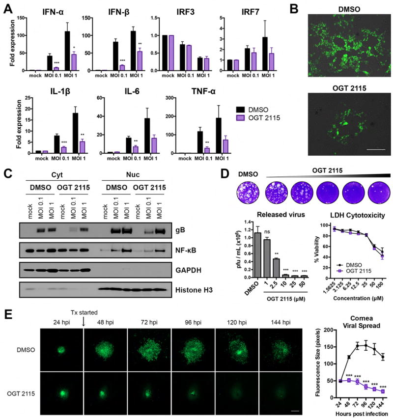
Herpes simplex virus-1 (HSV-1) causes lifelong recurrent pathologies without a cure. How infection by HSV-1 triggers disease processes, especially in the immune-privileged avascular human cornea, remains a major unresolved puzzle. It has been speculated that a cornea-resident molecule must tip the balance in favor of pro-inflammatory and pro-angiogenic conditions observed with herpetic, as well as non-herpetic, ailments of the cornea. Here, we demonstrate that heparanase (HPSE), a host enzyme, is the molecular trigger for multiple pathologies associated with HSV-1 infection. In human corneal epithelial cells, HSV-1 infection upregulates HPSE in a manner dependent on HSV-1 infected cell protein 34.5. HPSE then relocates to the nucleus to regulate cytokine production, inhibits wound closure, enhances viral spread, and thus generates a toxic local environment. Overall, our findings implicate activated HPSE as a driver of viral pathogenesis and call for further attention to this host protein in infection and other inflammatory disorders.

Keywords: cornea; cytokines; heparan sulfate; heparanase; herpes simplex virus; inflammation; interferon; ophthalmology; transcription factors; wound healing.


Conflict of interest statement

The authors declare that no conflict of interest exists.

Figures

Figure 1
Figure 1. Overexpression of constitutively active HPSE worsens disease in corneal HSV-1 infection
A, Schematic showing GS3-HPSE expression variant, in which the linker of the inactive 65-kDa form is replaced with a triple repeat of Gly-Ser to produce a constitutively active HPSE. Protein expression confirmed in HCE cell line with anti-HPSE PT-16673. B, Representative micrographs of mice 7 days post HSV-1 (McKrae 106 pfu) corneal infection. Scale bar, 1 mm. C, Clinical scores based on corneal opacity of mice in B. D, Representative micrographs of corneal surface of mice 14 days post HSV-1 (KOS 106 pfu) corneal infection. Scale bar, 1 mm. E–G, Analysis of ipsilateral draining (submandibular) lymph nodes of mice 7 days post HSV-1 (McKrae) infection. E, Representative flow cytometry data showing surface staining of F4/80 vs Gr-1, CD3 vs CD45, and CD4 vs CD8. Percentages of parent gates are labeled on each plot. F, Total numbers of cells in each mouse lymph node. G, Numbers of each cell type present in the draining lymph node. Asterisks denote a significant difference as determined by Student’s t-test; *P<0.05, **P<0.01, ***P<0.001, ns, not significant. See also Figure S1, S2, S6.
Figure 2
Figure 2. Active HPSE delays corneal wound healing in vivo and in vitro
A, Representative micrographs of in vivo corneal wound healing assay in murine corneas previously transfected with the GS3-HPSE or empty vector, in absence of infection. At specified timepoints post application of the circular wound, fluorescein was applied to highlight tissue damage (arrowheads), and corneas were imaged. Scale bar, 1 mm. B, Mean ± SEM of extent of wound healing in A. C, Representative micrographs of in vitro wound healing assay showing closure of HCE cellular defect over specified times after scratching. Before wound application, cells were transfected with empty vector, WT HPSE (not depicted) or GS3-HPSE. Scale bar, 50 μm. D, Quantification of extent of wound healing in D. Pixel distances between wound fronts in each panel were measured, and percentages of initial wound widths are plotted. E–F, The same procedure described in C and D was performed with the exception that HCE cells were infected with HSV-1 (KOS MOI 0.1) at the time of wound application. Asterisks denote a significant difference as determined by Student’s t-test; *P<0.05, **P<0.01, ***P<0.001, ns, not significant. See also Figure S3.
Figure 3
Figure 3. HPSE inhibits type I interferon signaling
A, Transcript copy numbers relative to GAPDH from HCE cells transfected with specified HPSE variants and HSV-1 infected. B, Luciferase assay results from HCE cells co-transfected with HPSE variants and pIFNb-luc, and infected for specified timepoints. C, Transcript copy numbers relative to GAPDH obtained as described in A. D, Representative western blot analysis of whole cell lysates of HCE cells transfected with specified HPSE variants in absence or presence of HSV-1. E, Densitometry analysis of D. F, Transcript copy numbers relative to GAPDH from wildtype and HPSE-knockout mouse embryonic fibroblasts in the absence or presence of HSV-1 for specified timepoints. G, Representative western blot analysis of whole cell lysates of wildtype and HPSE-knockout mouse embryonic fibroblasts in absence or presence of HSV-1 for specified timepoints. H, Densitometry analysis of G. All results are presented as mean ± SEM of three independent experiments (n=3). Asterisks denote a significant difference compared to EV, or WT MEF where applicable, for each timepoint, as determined by Student’s t-test; *P<0.05, **P<0.01, ***P<0.001, ns, not significant. See also Figure S3, S6.
Figure 4
Figure 4. HPSE translocates to nucleus upon infection and drives pro-inflammatory factor production and nuclear translocation of NF-κB
A, Transcript copy numbers of HCE cells transfected with specified HPSE variants, or transfected and HSV-1 infected for 24 h. B–C, Representative immunofluorescence micrographs of HCE cells mock treated or HSV-1 infected for 36 h. B, HPSE (red) and C, Heparan sulfate (green) are shown with respect to DAPI stain of nucleus (blue). Scale bar, 10 μm. D, Representative western blot analysis of cytoplasmic and nuclear extracts of HCE cells transfected with empty vector or GS3-HPSE. GAPDH and Histone H3 reflect cytoplasmic and nuclear content, respectively. E, Densitometry analysis expressed as fold change compared to EV mock samples, mean ± SEM of three independent experiments (n=3). F, Representative immunofluorescence micrographs of HCE cells transfected with empty vector or GS3-HPSE and HSV-1 infected for 24h. Nuclear translocation (arrowheads) of NF-κB p65 (green) is shown relative to DAPI (pseudocolored red). Scale bar, 10 μm. Asterisks denote a significant difference compared to EV for each timepoint, as determined by Student’s t-test; *P<0.05, **P<0.01, ***P<0.001, ns, not significant.
Figure 5
Figure 5. Viral upregulation of cellular HPSE can be driven by HSV-1 ICP34.5
A, Copy number of HPSE transcripts relative to GAPDH in HCE cells infected with HSV-1 (17) parental strain or 17Δγ34.5 mutant virus lacking ICP34.5 expression, or mock treated. B, Luciferase induction in HCE cells transfected with HPSE-luc plasmid and then infected for 24 h with HSV-1 (17) parental strain or 17Δγ34.5 mutant virus, or mock treated. C, Luciferase induction in HCE cells co-transfected with HPSE-luc plasmid and EV or γ34.5 plasmid, then assayed at specified timepoints. D, Representative immunofluorescence micrographs of porcine corneas infected for 24 h with HSV-1 (17) parental strain or 17Δγ34.5 mutant virus, then stained for HSV-1 (green), HPSE (red), and DAPI (blue). Antibody staining controls are presented in Figure S4. Scale bar, 100 μm. All plotted results are presented as mean ± SEM of three independent experiments (n=3). Asterisks denote a significant difference as determined by Student’s t-test; *P<0.05, **P<0.01, ***P<0.001, ns, not significant.
Figure 6
Figure 6. Pharmacological inhibition of HPSE by OGT 2115 blocks progression of HSV-1 infection
A, Transcript fold expression relative to DMSO mock, normalized to GAPDH, in HCE cells mock treated or infected with HSV-1 (KOS) for 24 h. B, Representative fluorescence micrographs of HCE cells infected with GFP-HSV-1 (K26-GFP MOI 0.1) in the presence of DMSO or OGT 2115. Scale bar, 50 μm. C, Representative western blot of cytoplasmic and nuclear extracts of HCE cells mock treated or infected for 24 h with HSV-1 (KOS) in the presence of DMSO or OGT 2115. GAPDH and Histone H3 reflect cytoplasmic and nuclear content, respectively. D, Representative plaque assay and quantification of virus released into supernatants of HCE cells infected with HSV-1 (KOS MOI 0.1) in the presence of DMSO or OGT 2115 at the specified concentrations. Lactate dehydrogenase (LDH) cytotoxicity assay results are shown at right. E, Representative fluorescence micrographs of ex vivo porcine corneas infected with GFP-HSV-1 (17-GFP) in the presence of DMSO or OGT 2115; quantification of fluorescence at right. Scale bar, 200 μm. All results are presented as mean ± SEM of three independent experiments (n=3). Asterisks denote a significant difference as determined by Student’s t-test; *P<0.05, **P<0.01, ***P<0.001, ns, not significant. See also Figure S5, S6.

References

    1. Agelidis AM, Shukla D. Cell entry mechanisms of HSV: what we have learned in recent years. Future Virol. 2015;10:1145–1154. - PMC - PubMed
    1. Alekseev O, Tran AH, Azizkhan-Clifford J. Ex Vivo Organotypic Corneal Model of Acute Epithelial Herpes Simplex Virus Type I Infection. JoVE 2012 - PMC - PubMed
    1. Aquino RS, Lee ES, Park PW. Diverse functions of glycosaminoglycans in infectious diseases. Prog Mol Biol Transl Sci. 2010;93:373–394. - PubMed
    1. Babu JS, Thomas J, Kanangat S, Morrison LA, Knipe DM, Rouse BT. Viral replication is required for induction of ocular immunopathology by herpes simplex virus. J Virol. 1996;70:101–107. - PMC - PubMed
    1. Bolovan CA, Sawtell NM, Thompson RL. ICP34.5 mutants of herpes simplex virus type 1 strain 17syn+ are attenuated for neurovirulence in mice and for replication in confluent primary mouse embryo cell cultures. J Virol. 1994;68:48–55. - PMC - PubMed

Publication types