. 2017 Jul 12;8(1):166.
doi: 10.1186/s13287-017-0616-0.

Isolation and characterization of equine endometrial mesenchymal stromal cells

Affiliations

Isolation and characterization of equine endometrial mesenchymal stromal cells

B Elisabeth Rink et al. Stem Cell Res Ther. .

Abstract

Background: Equine mesenchymal stromal/stem cells (MSCs) are most commonly harvested from bone marrow (BM) or adipose tissue, requiring the use of surgical procedures. By contrast, the uterus can be accessed nonsurgically, and may provide a more readily available cell source. While human endometrium is known to harbor mesenchymal precursor cells, MSCs have not been identified in equine endometrium. This study reports the isolation, culture, and characterization of MSCs from equine endometrium.

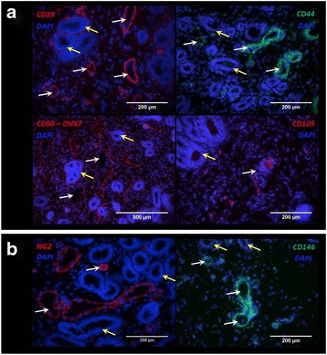
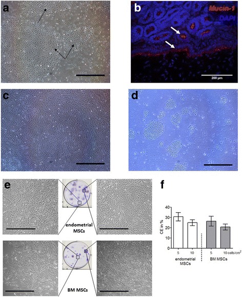
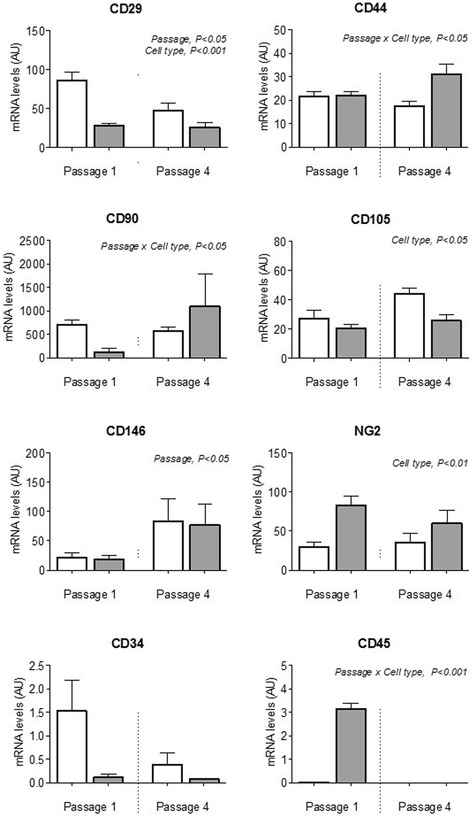
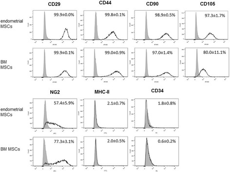
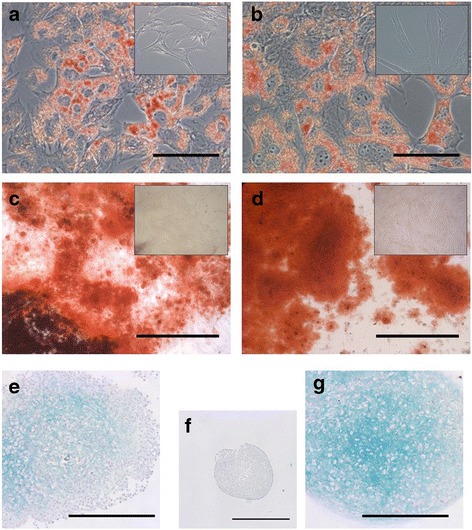
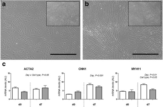
Methods: The presence of MSC and pericyte markers in endometrial sections was determined using immunohistochemistry. Stromal cells were harvested and cultured after separation of epithelial cells from endometrial fragments using Mucin-1-bound beads. For comparison, MSCs were also harvested from BM. The expression of surface markers in endometrial and BM-derived MSCs was characterized using flow cytometry and quantitative polymerase chain reaction. MSCs were differentiated in vitro into adipogenic, chondrogenic, osteogenic, and smooth muscle lineages.

Results: Typical markers of MSCs (CD29, CD44, CD90, and CD105) and pericytes (NG2 and CD146) were localized in the equine endometrium. Both endometrial and BM MSCs grew clonally and robustly expressed MSC and pericyte markers in culture while showing greatly reduced or negligible expression of hematopoietic markers (CD45, CD34) and MHC-II. Additionally, both endometrial and BM MSCs differentiated into adipogenic, osteogenic, and chondrogenic lineages in vitro, and endometrial MSCs had a distinct ability to undergo smooth muscle differentiation.

Conclusions: We have demonstrated for the first time the presence of cells in equine endometrium that fulfill the definition of MSCs. The equine endometrium may provide an alternative, easily accessible source of MSCs, not only for therapeutic regeneration of the uterus, but also for other tissues where MSCs from other sources are currently being used therapeutically.

Keywords: Endometrium; Equine; Horse; Mesenchymal stem cells.


Conflict of interest statement

Consent for publication

Not applicable.

Competing interests

The authors declare that they have no competing interests.

Publisher’s Note

Springer Nature remains neutral with regard to jurisdictional claims in published maps and institutional affiliations.

Figures

Fig. 1
Fig. 1
Immunohistochemistry of equine endometrial sections. Photomicrographs show localization of (a) MSC markers CD29, CD44, CD90, and CD105 and (b) perivascular markers NG2 and CD146 within the equine endometrium. DAPI was used to stain cell nuclei. Yellow arrows, endometrial glands; white arrows, blood vessels. DAPI 4′,6-diamidine-2′-phenylindole dihydrochloride (Color figure online)
Fig. 2
Fig. 2
Isolation and culture of MSCs. a Micrograph showing cells cultured directly following digestion of equine endometrium. Using this procedure, clusters of epithelial cells (black arrows) eventually outgrew stromal cells in culture. b Section of equine endometrium stained for Mucin-1, showing positive cells in epithelia and glands (white arrows). Cell nuclei stained with DAPI. c Micrograph of endometrial stromal cells in culture obtained after separation of epithelial cells (shown in d) using beads bound to Mucin-1. e Cell colonies produced after seeding of endometrial and BM MSCs at low densities. f Cloning efficiencies (CE) for endometrial MSCs (n = 6 horses) and BM MSCs (n = 3 horses) at two different seeding densities. Scale bars: 1 mm (a, ce). BM bone marrow, MSC mesenchymal stromal/stem cell (Color figure online)
Fig. 3
Fig. 3
Transcript levels (arbitrary units) of cell surface markers in cultured MSCs. Expression of MSC markers (CD29, CD44, CD90, CD105), perivascular markers (NG2, CD146), and hematopoietic markers (CD34, CD45) quantified by qPCR in endometrial MSCs (n = 6 horses, white bars) and BM MSCs (n = 3 horses, gray bars) in culture at passages 1 and 4. All results shown as mean ± SEM. Significant main effects (p < 0.05) of passage, cell type, and passage × cell type interaction obtained by two-way ANOVA are shown. AU arbitrary units
Fig. 4
Fig. 4
Flow cytometry analysis. Representative flow cytometry histograms with percentages of endometrial and BM MSCs (n = 6 and n = 3 horses, respectively) positive for different MSC, perivascular, and hematopoietic cell surface markers. Grey areas, signal from isotype controls; black lines, signal from the specific cell surface marker. BM bone marrow, MSC mesenchymal stromal/stem cell
Fig. 5
Fig. 5
Trilineage differentiation of endometrial MSCs (a, c, e) and BM MSCs (b, d, g). Representative images of endometrial and BM MSCs (n = 3 horses each) after differentiation and staining with Oil red O (a, b), Alizarin Red (c, d) and Alcian Blue/Nuclear Fast Red (eg) to assess differentiation into adipogenic, osteogenic, and chondrogenic lineages, respectively. Insets show nondifferentiated control cells (ad). Nondifferentiated control BM MSCs used in chondrogenic differentiation experiments shown in (f). Scale bars: 100 μm (a, b), 500 μm (c, d, e, g) and 1 mm (f)
Fig. 6
Fig. 6
Smooth muscle differentiation. Micrographs showing (a) endometrial MSCs (n = 3 horses) and (b) BM MSCs (n = 3 horses) induced to differentiate into smooth muscle for 7 days. Insets show noninduced control cells. Scale bars: 500 μm. c Expression of smooth muscle markers in endometrial MSCs (white bars) and BM MSCs (grey bars) before (d0) and on day 7 (d7) of differentiation. Results shown as mean ± SEM. Significant main effects (p < 0.05) of day, cell type, and day × cell type interaction obtained by two-way ANOVA are shown. AU arbitrary units

References

    1. Thomas ED, Lochte HL, Jr, Lu WC, Ferrebee JW. Intravenous infusion of bone marrow in patients receiving radiation and chemotherapy. N Engl J Med. 1957;257:491–6. doi: 10.1056/NEJM195709122571102. - DOI - PubMed
    1. Friedenstein AJ, Piatetzky S, II, Petrakova KV. Osteogenesis in transplants of bone marrow cells. J Embryol Exp Morphol. 1966;16:381–90. - PubMed
    1. Dominici M, Le Blanc K, Mueller I, Slaper-Cortenbach I, Marini F, Krause D, et al. Minimal criteria for defining multipotent mesenchymal stromal cells. The International Society for Cellular Therapy position statement. Cytotherapy. 2006;8:315–7. doi: 10.1080/14653240600855905. - DOI - PubMed
    1. Bourin P, Bunnell BA, Casteilla L, Dominici M, Katz AJ, March KL, et al. Stromal cells from the adipose tissue-derived stromal vascular fraction and culture expanded adipose tissue-derived stromal/stem cells: a joint statement of the International Federation for Adipose Therapeutics and Science (IFATS) and the International Society for Cellular Therapy (ISCT) Cytotherapy. 2013;15:641–8. doi: 10.1016/j.jcyt.2013.02.006. - DOI - PMC - PubMed
    1. Gargett CE, Schwab KE, Zillwood RM, Nguyen HPT, Wu D. Isolation and culture of epithelial progenitors and mesenchymal stem cells from human endometrium. Biol Reprod. 2009;80:1136–45. doi: 10.1095/biolreprod.108.075226. - DOI - PMC - PubMed

Substances