Review
. 2017 Jul 12;21(1):183.
doi: 10.1186/s13054-017-1750-x.

The future of mechanical ventilation: lessons from the present and the past

Affiliations
Review

The future of mechanical ventilation: lessons from the present and the past

Luciano Gattinoni et al. Crit Care. .

Abstract

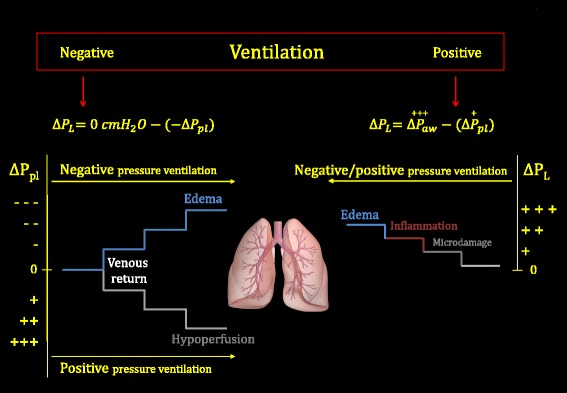
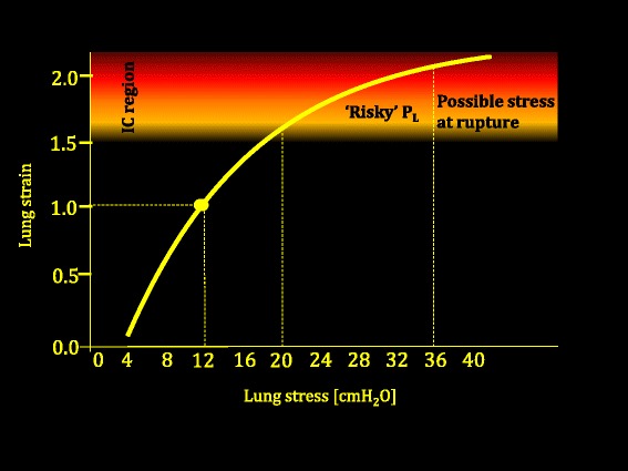
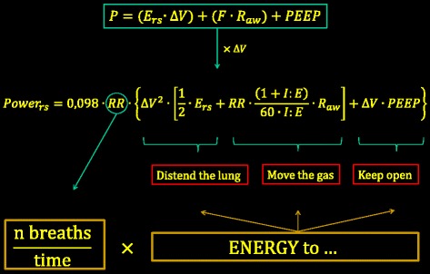
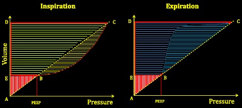
The adverse effects of mechanical ventilation in acute respiratory distress syndrome (ARDS) arise from two main causes: unphysiological increases of transpulmonary pressure and unphysiological increases/decreases of pleural pressure during positive or negative pressure ventilation. The transpulmonary pressure-related side effects primarily account for ventilator-induced lung injury (VILI) while the pleural pressure-related side effects primarily account for hemodynamic alterations. The changes of transpulmonary pressure and pleural pressure resulting from a given applied driving pressure depend on the relative elastances of the lung and chest wall. The term 'volutrauma' should refer to excessive strain, while 'barotrauma' should refer to excessive stress. Strains exceeding 1.5, corresponding to a stress above ~20 cmH2O in humans, are severely damaging in experimental animals. Apart from high tidal volumes and high transpulmonary pressures, the respiratory rate and inspiratory flow may also play roles in the genesis of VILI. We do not know which fraction of mortality is attributable to VILI with ventilation comparable to that reported in recent clinical practice surveys (tidal volume ~7.5 ml/kg, positive end-expiratory pressure (PEEP) ~8 cmH2O, rate ~20 bpm, associated mortality ~35%). Therefore, a more complete and individually personalized understanding of ARDS lung mechanics and its interaction with the ventilator is needed to improve future care. Knowledge of functional lung size would allow the quantitative estimation of strain. The determination of lung inhomogeneity/stress raisers would help assess local stresses; the measurement of lung recruitability would guide PEEP selection to optimize lung size and homogeneity. Finding a safety threshold for mechanical power, normalized to functional lung volume and tissue heterogeneity, may help precisely define the safety limits of ventilating the individual in question. When a mechanical ventilation set cannot be found to avoid an excessive risk of VILI, alternative methods (such as the artificial lung) should be considered.

Keywords: Acute respiratory distress syndrome; Extracorporeal membrane oxygenation; Mechanical power; Mechanical ventilation; Ventilator-induced lung injury.


Figures

Fig. 1
Fig. 1
Changes of transpulmonary pressure (∆P L) and of pleural pressure (∆P pl) during negative or positive pressure ventilation. Left: possible adverse consequences due to the progressive decrease or progressive increase of pleural pressure (∆P pl). The key variation is the increase or decrease of venous return, respectively. Right: sequence of possible damage when progressively increasing the transpulmonary pressure (∆P L). Either during negative pressure ventilation (here performed at baseline atmospheric pressure, i.e., 0 cmH2O) or during positive pressure ventilation, ∆P L is always positive. See text for details. ∆P aw change in airway pressure
Fig. 2
Fig. 2
Lung strain (tidal volume/FRC) as a function of lung stress (transpulmonary pressure). Data adapted from Agostoni and Hyatt [74]. As shown, the doubling of the FRC occurs at a transpulmonary pressure of 12 cmH2O (specific elastance). We arbitrarily indicated the ‘risky’ zone of P L as that which corresponds to lung strains exceeding 1.5 (based on experimental data [52]). P L transpulmonary pressure
Fig. 3
Fig. 3
Upper box: simplified equation of motion, showing that, at any given moment, the pressure in the respiratory system (P) above the relaxed volume equals the sum of the elastic pressure (elastance of the respiratory system E rs times change in lung volume), plus the pressure needed to move the gases (flow F times airway resistance), plus the pressure (if any) to keep the lung pressure above the atmospheric pressure at end expiration (PEEP). If each of these three components is multiplied by the tidal change in lung volume ∆V, the energy per breath is obtained. If multiplied by the respiratory rate, the corresponding power equation is obtained. 0.098 is the conversion factor from liters/cmH2O to Joules (J). I:E inspiratory–expiratory ratio, PEEP positive end-expiratory pressure, Power rs mechanical power to the respiratory system, RR respiratory rate, ∆V change of volume R aw airways resistances
Fig. 4
Fig. 4
Left: baseline energy (red hatched triangle ABE), on which the inspiratory energy associated with the tidal volume (area BCDE) is added. Yellow hatched area to the right of line BC represents the inspiratory dissipated energy needed to move the gas, to overcome surface tension forces, to make the extracellular sheets slide across one another (tissue resistances), and possibly to reinflate collapsed pulmonary units. Light green hatched area on the left of line BC defines the elastic energy (trapezoid EBCD) cyclically added to the respiratory system during inspiration. Total area included in the triangle ACD is the total energy level present in the respiratory system at end inspiration. Right: energy changes during expiration. Of the total energy accumulated at end inspiration (triangle ACD), the area of the trapezoid EBCD is the energy released during expiration. The fraction of energy included in the hysteresis area (light blue hatched area) is dissipated into the respiratory system, while the remaining area (dark blue hatched area) is energy dissipated into the atmosphere through the connecting circuit. Note that whatever maneuver (as controlled expiration) reduces the hysteresis area will reduce the energy dissipated into the respiratory system (potentially dangerous?). PEEP positive end-expiratory pressure (Color figure online)

References

    1. Vieillard-Baron A, et al. Experts’ opinion on management of hemodynamics in ARDS patients: focus on the effects of mechanical ventilation. Intensive Care Med. 2016;42(5):739–49. doi: 10.1007/s00134-016-4326-3. - DOI - PubMed
    1. Beitler JR, et al. Quantifying unintended exposure to high tidal volumes from breath stacking dyssynchrony in ARDS: the BREATHE criteria. Intensive Care Med. 2016;42(9):1427–36. doi: 10.1007/s00134-016-4423-3. - DOI - PMC - PubMed
    1. Files DC, Sanchez MA, Morris PE. A conceptual framework: the early and late phases of skeletal muscle dysfunction in the acute respiratory distress syndrome. Crit Care. 2015;19:266. doi: 10.1186/s13054-015-0979-5. - DOI - PMC - PubMed
    1. Petrof BJ, Hussain SN. Ventilator-induced diaphragmatic dysfunction: what have we learned? Curr Opin Crit Care. 2016;22(1):67–72. doi: 10.1097/MCC.0000000000000272. - DOI - PubMed
    1. American Thoracic Society. Infectious Diseases Society of America Guidelines for the management of adults with hospital-acquired, ventilator-associated, and healthcare-associated pneumonia. Am J Respir Crit Care Med. 2005;171(4):388–416. doi: 10.1164/rccm.200405-644ST. - DOI - PubMed

MeSH terms