. 2017 Sep 1;313(3):C314-C326.
doi: 10.1152/ajpcell.00229.2016. Epub 2017 Jul 12.

Mechanism of action of the anti-inflammatory connexin43 mimetic peptide JM2

Affiliations

Mechanism of action of the anti-inflammatory connexin43 mimetic peptide JM2

J Matthew Rhett et al. Am J Physiol Cell Physiol. .

Abstract

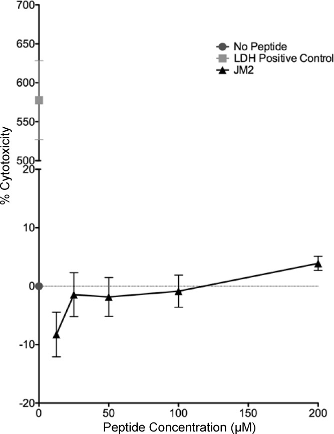
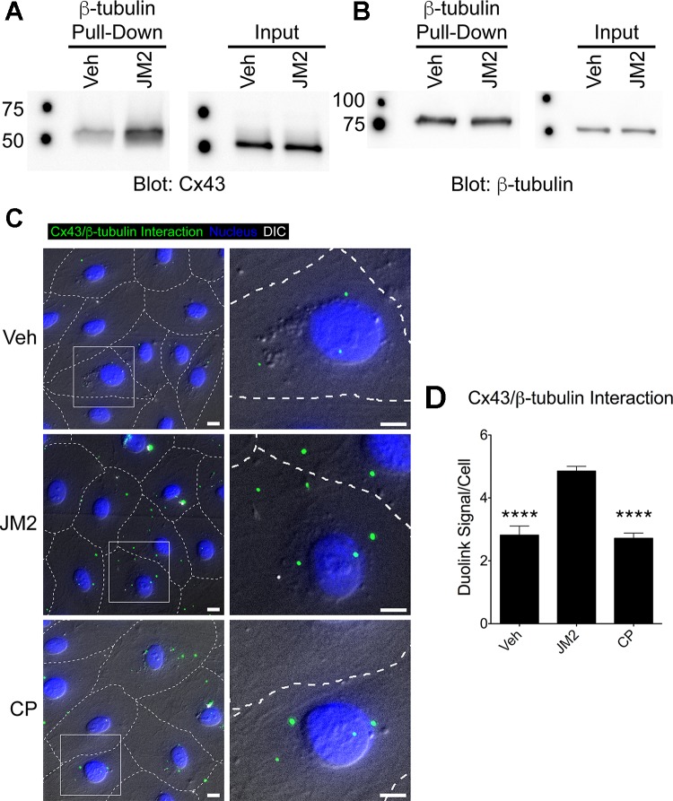
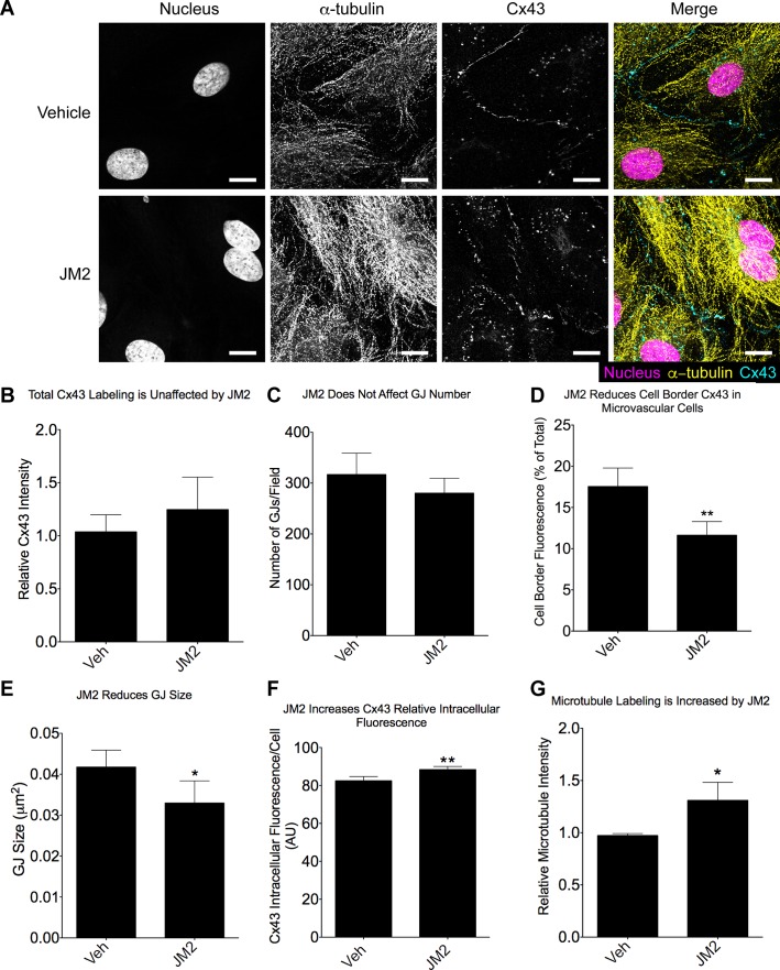
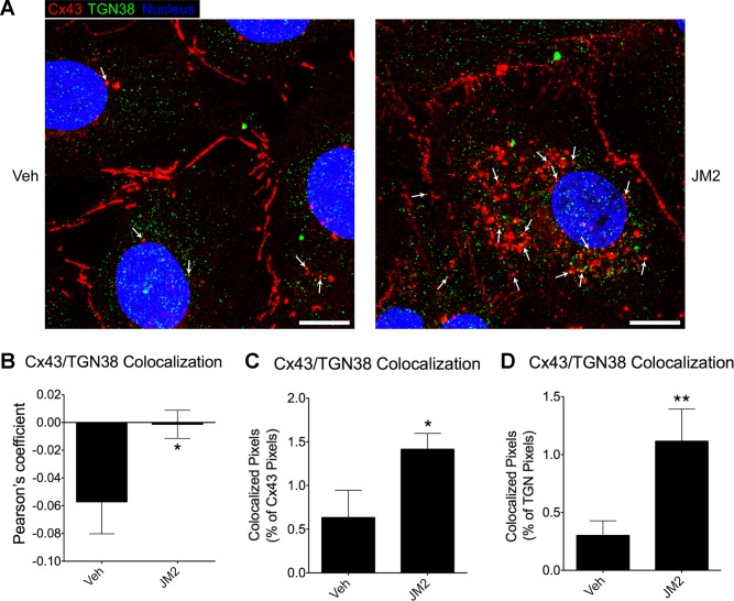
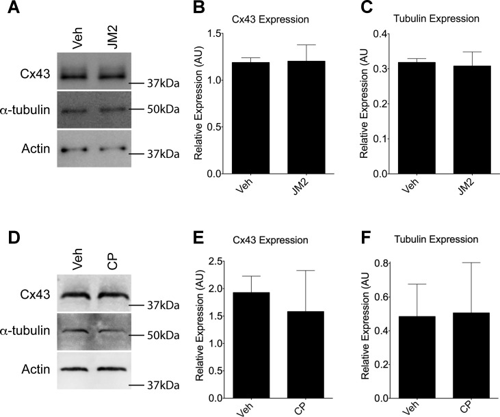
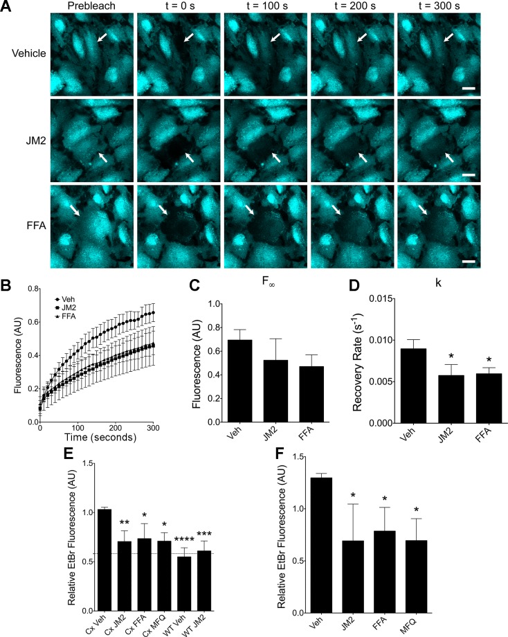
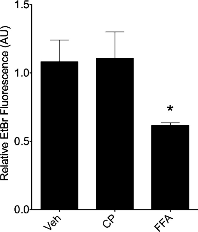
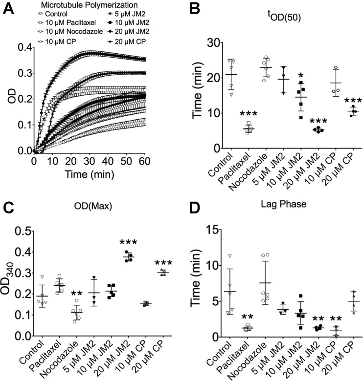
Connexin-based therapeutics have shown the potential for therapeutic efficacy in improving wound healing. Our previous work demonstrated that the connexin43 (Cx43) mimetic peptide juxtamembrane 2 (JM2) reduced the acute inflammatory response to a submuscular implant model by inhibiting purinergic signaling. Given the prospective application in improving tissue-engineered construct tolerance that these results indicated, we sought to determine the mechanism of action for JM2 in the present study. Using confocal microscopy, a gap-FRAP cell communication assay, and an ethidium bromide uptake assay of hemichannel function we found that the peptide reduced cell surface Cx43 levels, Cx43 gap junction (GJ) size, GJ communication, and hemichannel activity. JM2 is based on the sequence of the Cx43 microtubule binding domain, and microtubules have a confirmed role in intracellular trafficking of Cx43 vesicles. Therefore, we tested the effect of JM2 on Cx43-microtubule interaction and microtubule polymerization. We found that JM2 enhanced Cx43-microtubule interaction and that microtubule polymerization was significantly enhanced. Taken together, these data suggest that JM2 inhibits trafficking of Cx43 to the cell surface by promoting irrelevant microtubule polymerization and thereby reduces the number of hemichannels in the plasma membrane available to participate in proinflammatory purinergic signaling. Importantly, this work indicates that JM2 may have therapeutic value in the treatment of proliferative diseases such as cancer. We conclude that the targeted action of JM2 on Cx43 channels may improve the tolerance of implanted tissue-engineered constructs against the innate inflammatory response.

Keywords: cancer; connexin43; gap junction; hemichannel; inflammation.


Figures

Fig. 1.
Fig. 1.
Juxtamembrane 2 (JM2) is not cytotoxic. Human microvascular endothelial cells (HMVECs) were treated with JM2 at 12.5, 25, 50, 100, and 200 μM concentrations for 2 h at 37°C, 5% CO2. The media were then sampled for lactate dehydrogenase (LDH) as a marker for cell death. No significant differences were observed between control (No Peptide) and any concentration of JM2. n = 3, with each n representing the average of 3 replicates; error bars represent SE.
Fig. 2.
Fig. 2.
JM2 specifically enhances connexin43 (Cx43)-β-tubulin interaction. A and B: Cx43-HeLa cell lysates were incubated with β-tubulin-glutathione S-transferase (GST) bound to glutathione-Sepharose beads plus vehicle (H2O; Veh) or 25 μM JM2. Eluted proteins were analyzed by immunoblotting for Cx43 (A) or β-tubulin (B). β-Tubulin pulled down Cx43 in control conditions (vehicle), and this was increased in the presence of JM2 (A, “β-tubulin Pull-Down”), indicating that JM2 specifically enhances the interaction between Cx43 and β-tubulin. This was supported by the observation that the input amounts of Cx43 and β-tubulin subjected to pull-down were equivalent between Veh and JM2 treatments (A and B “Input” blots, respectively) and that JM2 did not affect the amount of β-tubulin pulled down by a β-tubulin antibody (B, “β-tubulin Pull-Down”). C: HMVECs were treated for 2 h with either vehicle (H2O; “Veh”), 50 μM JM2, or 50 μM control peptide (CP). Cells were fixed and labeled for Cx43-β-tubulin interaction (green), and the nucleus (blue). Differential interference contrast (DIC; gray scale) was used to approximate cell-cell borders (dashed lines). Boxed regions indicate the location of the enlarged insets. Scale bar = 10 μm. D: Cx43-β-tubulin interaction was quantified as the number of Duolink signals detected per cell. The number of signals detected in JM2-treated cells was significantly greater than in vehicle (Veh) or control peptide (CP). n = 3, with each n representing the average of 5 replicates; error bars represent SD. Nonlinear level adjustments were applied to Western blot images to enhance visibility. ****P < 0.001.
Fig. 3.
Fig. 3.
JM2 decreases gap junction (GJ) size while increasing labeling for microtubules. HMVECs were treated for 2 h with either vehicle (H2O; “Veh”) or 50 μM JM2. A: cells were fixed and labeled for Cx43 (cyan), α-tubulin (yellow), and the nucleus (magenta); Scale bar = 10 μm. B: total Cx43 fluorescence was unaffected by JM2. C and D: similarly, GJ number was measured and found to be unaffected by JM2 (C), while there was a significant decrease in the level of cell border-associated Cx43 fluorescence (D). E: consistent with the reduction in cell border Cx43 fluorescence, GJ size was reduced. F: concomitantly, the level of intracellular Cx43 fluorescence was significantly increased in JM2-treated cells. G: microtubule fluorescence was also significantly increased by JM2 treatment. For all graphs, n = 3, with each n representing the average of 5 replicates; error bars represent SD. *P < 0.05, **P < 0.01.
Fig. 4.
Fig. 4.
JM2 increases the accumulation of Cx43-containing secretory vesicles. HMVECs were treated for 2 h with either vehicle (H2O; “Veh”) or 50 μM JM2. A: cells were fixed and labeled for Cx43 (red), the trans-Golgi network (TGN) protein TGN38 (green), and the nucleus (blue). Arrows indicate a number of puncta with Cx43 TGN38 colocalization that are likely secretory vesicles; Scale bar = 10 μm. BD: colocalization of Cx43 and TGN38 was significantly increased by JM2 as assessed by Pearson’s coefficient (B), the amount of colocalized pixels as a % of total Cx43 pixels (C), and the amount of colocalized pixels as a % of total TGN38 pixels (D); n = 3, with each n representing the average of 3 replicates; error bars represent SD. *P < 0.05, **P < 0.01.
Fig. 5.
Fig. 5.
Cx43 and tubulin expression are unaffected by JM2. A and D: Cx43, α-tubulin, and actin immunoblots of HMVEC lysates from cultures treated with vehicle (H2O; “Veh”), 50 μM JM2 (A), or 50 μM control peptide (D). B, C, E, and F: densitometry was performed, and Cx43 (B and E) and α-tubulin (C and F) levels were normalized to actin for JM2 (B and C) and control peptide (E and F) treated cells. For all graphs, n = 3; error bars represent SD. No significant changes were observed. Nonlinear level adjustments were applied to Western blot images to enhance visibility. AU, arbitrary units.
Fig. 6.
Fig. 6.
JM2 treatment inhibits both gap junction intercellular communication and hemichannel function. A: HMVECs were treated with either vehicle (H2O and EtOH), 50 μM JM2, or 50 μM flufenamic acid (FFA) for 2 h, and calcein-AM was loaded during the final 30 min. Selected cells (arrows) were then photobleached by high-intensity laser light and recovery was monitored. Scale bar = 20 μm. B: average fluorescence readings were plotted over time. n = 3, with each n representing the average of 3 replicates; error bars represent SE. C and D: nonlinear regression to an exponential decay function was used to determine the maximum predicted recovery (F; C) and the recovery rate constant (k; D). Error bars represent SD. E: connexin43 (Cx43) and and wild-type (WT) HeLa cells were treated with either vehicle (H2O and EtOH; “Veh”), 50 μM JM2, 50 μM FFA, or 5 μM mefloquine (MFQ) for 2 h, then exposed to ethidium bromide (EtBr) for 15 min. Cells were fixed and imaged, and the quantified relative fluorescence is displayed. Dashed line indicates the level of autofluorescence in Cx43-HeLa cells. F: HMVECs were treated, imaged, and quantified as in E. For graphs in E and F, n = 4, with each n representing the average of 5 replicates; error bars represent SD. *P < 0.05, **P < 0.01, ***P< 0.001, ****P < 0.001.
Fig. 7.
Fig. 7.
A control peptide does not affect hemichannel function. HMVECs were treated with either vehicle (H2O and EtOH; “Veh”), 50 μM control peptide (CP), or 50 μM FFA for 2 h, then exposed to EtBr for 15 min. Cells were fixed and imaged, and the quantified relative fluorescence is displayed. No significant difference was observed between vehicle control and control peptide treatment conditions; n = 3, with each n representing the average of 5 replicates; error bars represent SD. *P < 0.05.
Fig. 8.
Fig. 8.
The Cx43 tubulin binding domain induces microtubule polymerization. A: microtubule polymerization was spectrophotometrically monitored by reading absorbance at 340 nm over a period of 1 h in control, 5, 10, and 20 μM JM2, 10 and 20 μM control peptide (CP), 10 μM paclitaxel, and 10 μM nocodazole treatment conditions. Error bars represent SE. B: time (t) to 50% maximum OD340 (tOD(50)) was determined as a proxy for reaction rate. C: maximum OD was measured to quantify the peak conversion of tubulin to microtubules. D: the duration of the lag phase was used as a measure of the nucleation phase. For B, C, D, and E, n ≥ 3; error bars represent SD. *P < 0.05, **P < 0.01, ***P< 0.001.

References

    1. Abbaci M, Barberi-Heyob M, Blondel W, Guillemin F, Didelon J. Advantages and limitations of commonly used methods to assay the molecular permeability of gap junctional intercellular communication. Biotechniques 45: 33–62, 2008. doi:10.2144/000112810. - DOI - PubMed
    1. Bruzzone R, Barbe MT, Jakob NJ, Monyer H. Pharmacological properties of homomeric and heteromeric pannexin hemichannels expressed in Xenopus oocytes. J Neurochem 92: 1033–1043, 2005. doi:10.1111/j.1471-4159.2004.02947.x. - DOI - PubMed
    1. Burdzinska A, Gala K, Kowalewski C, Zagozdzon R, Gajewski Z, Pa̦czek L. Dynamics of acute local inflammatory response after autologous transplantation of muscle-derived cells into the skeletal muscle. Mediators Inflamm 2014: 482352, 2014. doi:10.1155/2014/482352. - DOI - PMC - PubMed
    1. Burnstock G. Introductory overview of purinergic signalling. Front Biosci (Elite Ed) E3: 896–900, 2011. doi:10.2741/e298. - DOI - PubMed
    1. Calder BW, Rhett MJ, Bainbridge H, Fann SA, Gourdie RG, Yost MJ. Inhibition of connexin 43 hemichannel-mediated ATP release attenuates early inflammation during the foreign body response. Tissue Eng Part A 21: 1752–1762, 2015. doi:10.1089/ten.tea.2014.0651. - DOI - PMC - PubMed

MeSH terms