Review
. 2017 Oct 1;118(4):2181-2193.
doi: 10.1152/jn.00283.2017. Epub 2017 Jul 12.

Measuring and quantifying skin sympathetic nervous system activity in humans

Affiliations
Review

Measuring and quantifying skin sympathetic nervous system activity in humans

Jody L Greaney et al. J Neurophysiol. .

Abstract

Development of the technique of microneurography has substantially increased our understanding of the function of the sympathetic nervous system (SNS) in health and in disease. The ability to directly record signals from peripheral autonomic nerves in conscious humans allows for qualitative and quantitative characterization of SNS responses to specific stimuli and over time. Furthermore, distinct neural outflow to muscle (MSNA) and skin (SSNA) can be delineated. However, there are limitations and caveats to the use of microneurography, measurement criteria, and signal analysis and interpretation. MSNA recordings have a longer history and are considered relatively more straightforward from a measurement and analysis perspective. This brief review provides an overview of the development of the technique as used to measure SSNA. The focus is on the utility of measuring sympathetic activity directed to the skin, the unique issues related to analyzing and quantifying multiunit SSNA, and the challenges related to its interpretation.

Keywords: MSNA; SSNA; cutaneous blood flow; microneurography; thermoregulation; vasoconstriction; vasodilation.


Figures

Fig. 1.
Fig. 1.
Original recordings of muscle sympathetic nerve activity (MSNA; A) and skin sympathetic nerve activity (SSNA; B) depicting the distinct burst morphology and temporal patterns used by microneurographers to distinguish between multiunit efferent MSNA and SSNA recordings. Arterial blood pressure (BP; Finometer) and heart rate (electrocardiogram) are shown for reference.
Fig. 2.
Fig. 2.
Representation of the various techniques employed for the normalization of baseline SSNA in multiunit recordings. Normalization to the mean strength of spontaneous activity at rest, in the absence of any stimulus, involves assigning an arbitrary value to a baseline segment of SSNA, represented by setting the integration of the green-shaded area to 100. Normalization to the highest burst of spontaneous activity at rest is represented by the hashed red box, in which the largest burst is assigned a value of 100. In the last approach, a baseline segment containing a long nonbursting period is set to 0 arbitrary units, depicted by the blue box. Each of these normalization procedures allows for the within-subject assessment of SSNA responsiveness relative to a given stimulus.
Fig. 3.
Fig. 3.
Schematic diagram depicting thermoregulatory control of skin sympathetic nerve activity (SSNA) to skin effectors. The thermal input signal (I) is a weighted function of core (Tc) and mean skin (Tsk) temperatures (i.e., I = α1·Tc + α2·Tsk + b), where the ratio of α12 is ~9:1 for whole body heating and 7:3 for whole body cooling. Signal input is also modified by local skin temperature and rate of change parameters (not shown), as well as nonthermoregulatory inputs. The input signal I is compared with a “set point” (I0) in the preoptic area of the hypothalamus (POA), and the error signal (I − I0) determines the output efferent SSNA. Note that set point is conceptual only and not an actual temperature. SSNA determines the onset and gain characteristics of the skin thermoregulatory effector organs, controlling vasoconstriction and piloerection in the cold and vasodilation and sweating in the heat. DRG, dorsal root ganglion; IML, intermediolateral nucleus; RMR, rostral medullary raphe.
Fig. 4.
Fig. 4.
Original recording of multiunit skin sympathetic nerve activity (SSNA) at baseline and during whole body cooling (A). During cooling, vasoconstrictor bursts are wider and have a longer duration. For comparison, an original recording of multiunit SSNA at thermoneutrality and during whole body heating is also presented (B). Bursts of SSNA become more narrow and begin to appear morphologically similar to bursts of muscle sympathetic nerve activity. Tsk, skin temperature; Tes, esophageal temperature.
Fig. 5.
Fig. 5.
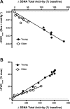
There was a significant relation between the increase in skin sympathetic nervous system activity (SSNA) and the reduction in cutaneous vascular conductance (CVC) during whole body cooling (i.e., reduction in mean skin temperature from 34.0° to 30.5°C; A) and whole body heating (i.e., increase in core temperature of 1.0°C; B) in both young (●) and healthy older adults (○). In healthy older adults, blunted cooling-induced increases in SSNA were linearly related to impaired reflex cutaneous vasoconstriction. The slope of the ∆SSNA:∆CVC relation was not different between groups, suggesting that the relative inability of older adults to decrease skin blood flow during whole body cooling is not reflective of diminished sensitivity of the neural reflex response but is instead indicative of age-related reductions in the range of efferent SSNA responsiveness to cooling. Similarly, blunted heating-induced increases in SSNA were linearly related to impaired reflex cutaneous vasodilation in healthy older adults. However, in contrast to cooling, the slope of the ∆SSNA:∆CVC relation during heating was also reduced in healthy older adults, suggesting that healthy aging is characterized by a reduced reflex vasodilatory response to increased SSNA. [Adapted from Greaney et al. (2015b) and Stanhewicz et al. (2016).]

References

    1. Aguayo AJ, Bray GM, Terry LC, Sweezey E. Three dimensional analysis of unmyelinated fibers in normal and pathologic autonomic nerves. J Neuropathol Exp Neurol 35: 136–151, 1976. doi:10.1097/00005072-197603000-00002. - DOI - PubMed
    1. Bernjak A, Cui J, Iwase S, Mano T, Stefanovska A, Eckberg DL. Human sympathetic outflows to skin and muscle target organs fluctuate concordantly over a wide range of time-varying frequencies. J Physiol 590: 363–375, 2012. doi:10.1113/jphysiol.2011.214528. - DOI - PMC - PubMed
    1. Bini G, Hagbarth KE, Hynninen P, Wallin BG. Regional similarities and differences in thermoregulatory vaso- and sudomotor tone. J Physiol 306: 553–565, 1980a. doi:10.1113/jphysiol.1980.sp013414. - DOI - PMC - PubMed
    1. Bini G, Hagbarth KE, Hynninen P, Wallin BG. Thermoregulatory and rhythm-generating mechanisms governing the sudomotor and vasoconstrictor outflow in human cutaneous nerves. J Physiol 306: 537–552, 1980b. doi:10.1113/jphysiol.1980.sp013413. - DOI - PMC - PubMed
    1. Blumberg H, Wallin BG. Direct evidence of neurally mediated vasodilatation in hairy skin of the human foot. J Physiol 382: 105–121, 1987. doi:10.1113/jphysiol.1987.sp016358. - DOI - PMC - PubMed

Publication types

LinkOut - more resources