. 2017 Jul 12;7(1):5210.
doi: 10.1038/s41598-017-05549-w.

Secretome of in vitro cultured human embryos contains extracellular vesicles that are uptaken by the maternal side

Affiliations

Secretome of in vitro cultured human embryos contains extracellular vesicles that are uptaken by the maternal side

Elisa Giacomini et al. Sci Rep. .

Abstract

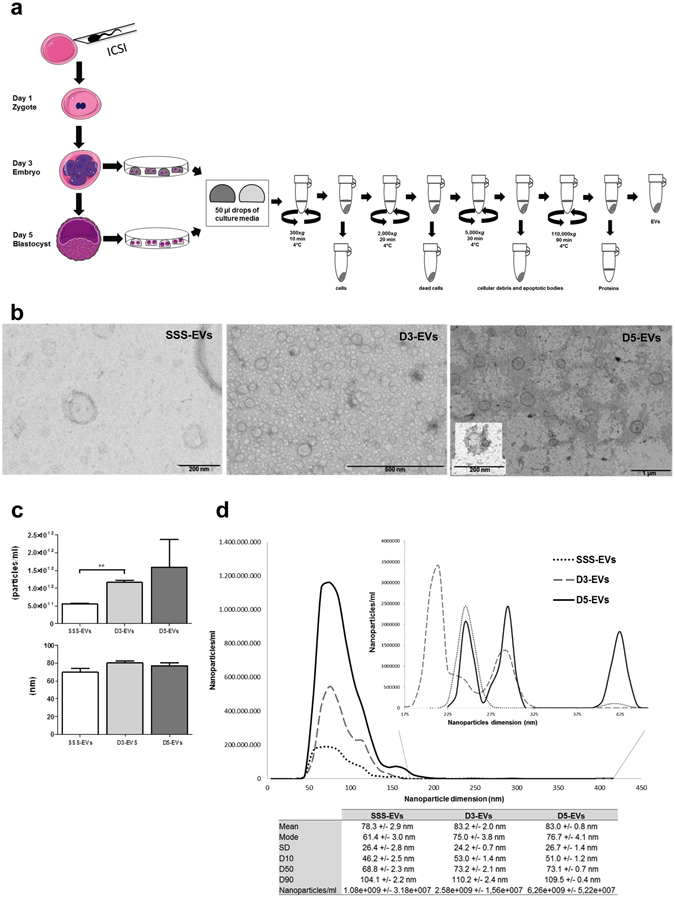
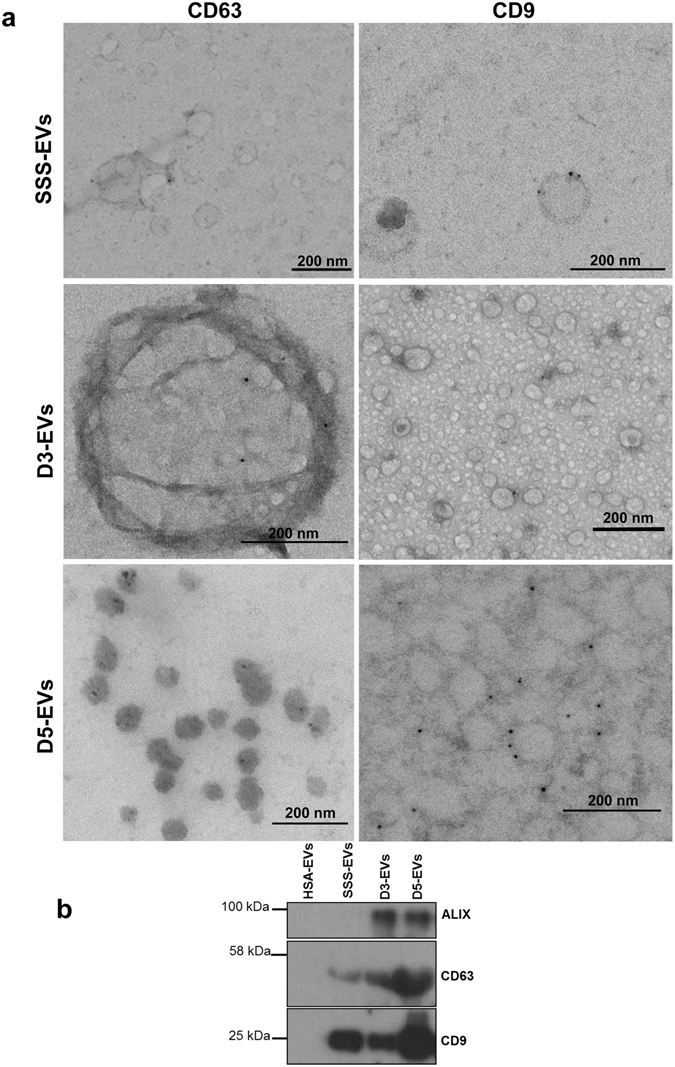
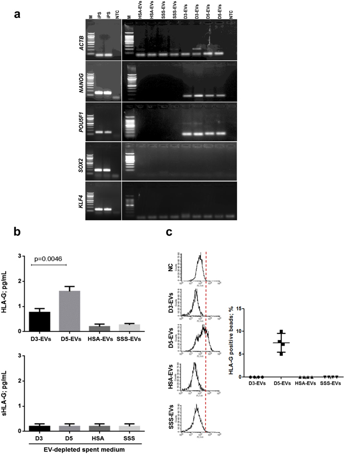
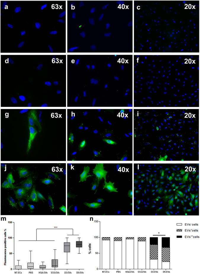
Communication between embryo and maternal endometrium occurs during a specific time frame in which implantation is possible. Here we demonstrate for the first time that conditioned media from non-manipulated human embryos cultured in vitro for 3 days or up to the blastocyst stage contain extracellular vesicles (EVs) with a diameter of 50 to 200 nm and bearing the traditional microvesicle and exosome marker proteins CD63, CD9 and ALIX. The embryonic origin of these EVs has been confirmed by the presence of stemness gene transcripts and their enrichment in the non-classical HLA-G protein. NANOG and POU5F1 transcripts were shown to be contained in vesicles deriving from embryos at different stages of development. In line with a higher detection rate of the HLA-G protein in blastocysts compared to cleavage stage embryos, a significantly higher amount of HLA-G was found in vesicles accumulated in spent media from day 3 to day 5 of development compared to those isolated from the earlier stage. Uptake of dye-labeled embryo-derived EVs by human primary endometrial epithelial and stromal cells was also demonstrated with a fluorescence intensity signal significantly higher for cells treated with vesicles derived from blastocysts. Based on these findings, EV exchange may be suggested as an emerging way of communication at the maternal-fetal interface.


Conflict of interest statement

The authors declare that they have no competing interests.

Figures

Figure 1
Figure 1
Day 3 and day 5 embryos release a heterogeneous population of EVs. (a) Schematic overview of the isolation protocol used to obtain EVs from embryo spent media accumulated from day 1 to day 3 (D3-EVs) or from day 3 to day 5 (D5-EVs) of development. (b) Fresh D3-EVs or D5-EVs isolated from six embryos were negatively stained with uranyl acetate and visualized by TEM. EVs isolated from SSS-supplemented medium (SSS-EVs) are also shown (left panel). Data shown are representative of 3 independent experiments. (c) Concentration defined as particle/ml (upper panel) and size distribution (lower panel) profiles were determined by NTA using 50 µl of spent media from six day 3 or day 5 embryos. Size and number of SSS-EVs are also shown. Results are expressed as mean ± SEM, n = 4 (**p = 0.001, D3-EVs vs SSS-EVs). (d) A representative tracking analysis plot of SSS-EVs (dilution 1:500) and of D3-EVs and D5-EVs (dilution 1:500). To better visualize peaks, a different scale was used for nanoparticles of diameter higher than 175 nm. The mode, mean value, standard deviation of size and concentration for each sample are provided. The value D50 represents the median size. Similarly, 90 percent of the distribution lies below the D90 value, and 10 percent of the population lies below the D10 value.
Figure 2
Figure 2
Marker characterization of EVs released by day 3 and day 5 embryos. (a) EVs isolated from a single drop of spent media from six embryos at day 3 (D3-EVs) or day 5 (D5-EVs) developmental stage were negatively stained with uranyl acetate and visualized by TEM after immunogold labelling with anti-CD63 and anti-CD9 antibodies. Transmission electron microscopy images of EVs from SSS-supplemented medium (SSS-EVs) after immunogold labelling with anti-CD9 and anti-CD63 is also shown (upper panels). Black dots indicate membrane-associated gold. Data shown are representative of 3 independent experiments. (b) Ten µg of proteins isolated from D3-EVs and D5-EVs and from an equal volume of fresh 10% SSS- or 5% HSA-supplemented media were analyzed for the presence of molecular markers of microvesicles and of exosomes by western blotting. Data shown are representative of 3 independent experiments.
Figure 3
Figure 3
EVs isolated from day 3 and day 5 embryos contain embryonic specific molecules. (a) Five ng of EVs-derived amplified cDNA from a pool of spent media from 50 day 3 (D3-EVs) or day 5 (D5-EVs) embryos were subjected to touch down PCR in order to assess presence of pluripotency-related gene transcripts. Gel electrophoresis of PCR products is shown. iPS cDNA was used as a positive control and EVs derived from fresh media conditioned with 10% of SSS or 5% of HSA were used as negative controls (HSA-EVs and SSS-EVs). No template controls (NTC) were also added to distinguish the primer dimer formation. Three independent experiments have been performed and two different samples per condition are shown. M = 100 bp molecular ladder. (b) HLA-G levels in EVs (upper panel) from a pool of spent media of 50 embryos in day 3 (D3-EVs) or day 5 (D5-EVs) of development were evaluated by Bio-Plex assay. Equal volumes of fresh media conditioned with 10% of SSS (SSS-EVs or SSS) or 5% of HSA (HSA-EVs or HSA) were used as negative controls. Results are expressed as mean ± SEM, n = 6 (p < 0.0046, D5-EVs vs D3-EVs). Soluble HLA-G levels have been also evaluated in EV-depleted spent culture media from the same samples by Bio-Plex assay, n = 3 (lower panel). (c) Flow cytometry analysis was performed to confirm the levels of HLA-G in EVs. The same analysis was performed in both EVs and in transfected HeLa-G5 cell culture supernatants used as positive control (not shown) and in HeLa wild-type cell culture supernatants as negative control (NC). The samples were analyzed in duplicates. Results are expressed as mean ± SEM, n = 4.
Figure 4
Figure 4
Primary ECs can incorporate embryo-derived EVs. Representative images of primary ECs treated for 4 hours with 10 µg/ml of Vybrant DiO-labelled EVs derived from 5% HSA-supplemented fresh medium (HSA-EVs) (ac), from 10% SSS-supplemented fresh media (SSS-EVs) (df) and from conditioned media of day 3 embryos (D3-EVs) (gi) and of day 5 embryos (D5-EVs) (jl). Blue = nuclei. Box plot of percentage of dye-positive cells (EV+ plus EV++cells). Block lines within boxes represent median value and the whiskers indicate the minimum and the maximum [N = 3, ***P < 0.0001, D3-EVs/D5-EVs vs all negative controls (PBS, HSA-EVs and SSS-EVs)] (m); engrafted distributions of fluorescence intensity among the different EV species used for primary cell treatment (*P = 0.01, D3-EVs vs D5-EVs) (n).

References

    1. Steptoe PC, Edwards RG. Reimplantation of a human embryo with subsequent tubal pregnancy. Lancet. 1976;1(7965):880. doi: 10.1016/S0140-6736(76)92096-1. - DOI - PubMed
    1. Wang J, Sauer MV. In vitro fertilization (IVF): a review of 3 decades of clinical innovation and technological advancement. Ther. Clin. Risk. Manag. 2006;2(4):355–64. doi: 10.2147/tcrm.2006.2.4.355. - DOI - PMC - PubMed
    1. Wade JJ, MacLachlan V, Kovacs G. The success rate of IVF has significantly improved over the last decade. Aust. N. Z. J. Obstet. Gynaecol. 2015;55(5):473–6. doi: 10.1111/ajo.12356. - DOI - PubMed
    1. Zhang S, et al. Physiological and molecular determinants of embryo implantation. Mol. Aspects Med. 2013;34(5):939–80. doi: 10.1016/j.mam.2012.12.011. - DOI - PMC - PubMed
    1. Cha J, Sun X, Dey SK. Mechanisms of implantation: strategies for successful pregnancy. Nat. Med. 2012;18(12):1754–67. doi: 10.1038/nm.3012. - DOI - PMC - PubMed

Publication types