. 2017 Jul 12;7(1):5170.
doi: 10.1038/s41598-017-05500-z.

Oncolytic measles virus enhances antitumour responses of adoptive CD8+NKG2D+ cells in hepatocellular carcinoma treatment

Affiliations

Oncolytic measles virus enhances antitumour responses of adoptive CD8+NKG2D+ cells in hepatocellular carcinoma treatment

Aiping Chen et al. Sci Rep. .

Abstract

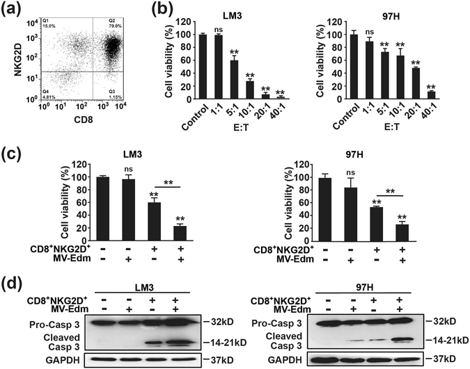
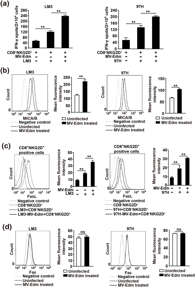
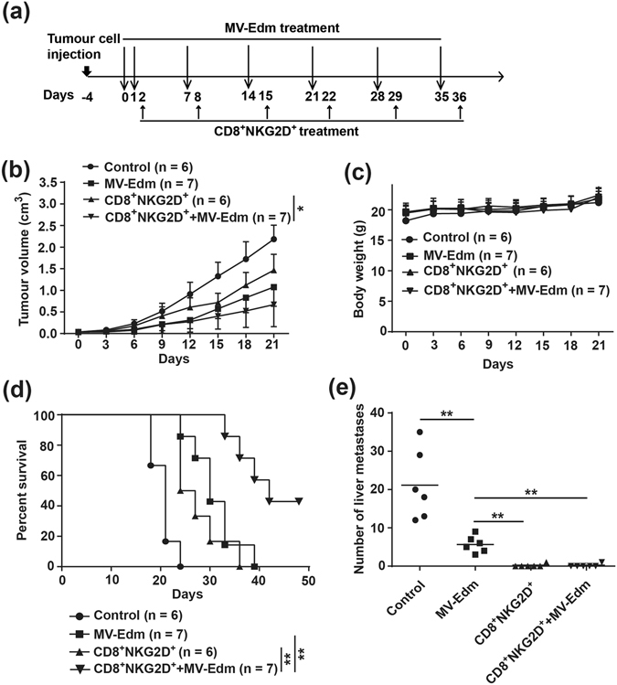
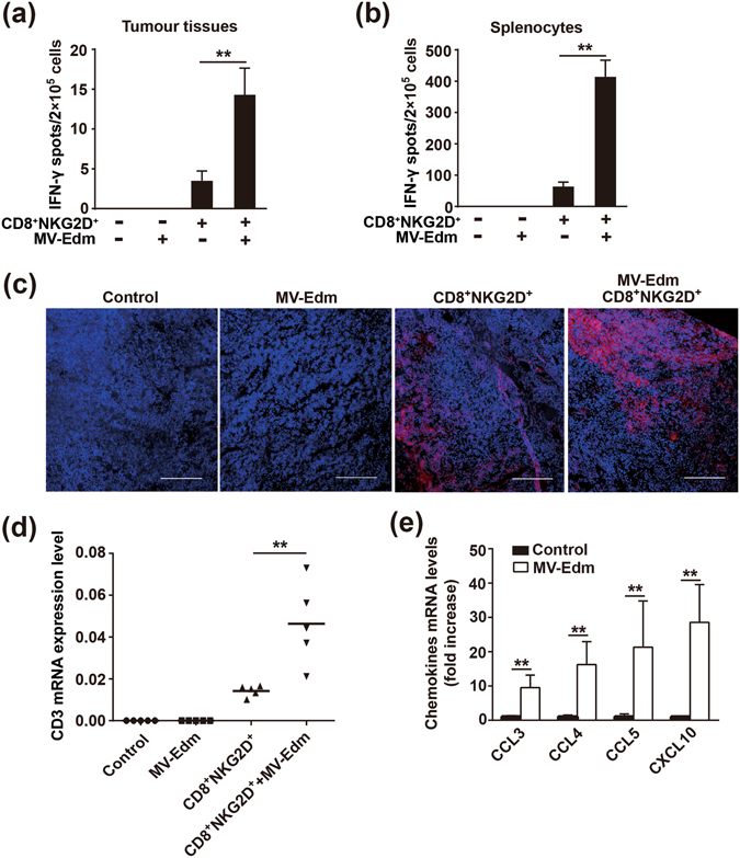
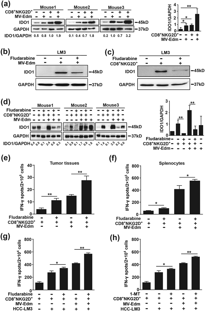
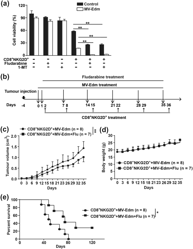
There is an urgent need for novel effective treatment for hepatocellular carcinoma (HCC). Oncolytic viruses (OVs) not only directly lyse malignant cells, but also induce potent antitumour immune responses. The potency and precise mechanisms of antitumour immune activation by attenuated measles virus remain unclear. In this study, we investigated the potency of the measles virus vaccine strain Edmonston (MV-Edm) in improving adoptive CD8+NKG2D+ cells for HCC treatment. We show that MV-Edm-infected HCC enhanced the antitumour activity of CD8+NKG2D+ cells, mediated by at least three distinct mechanisms. First, MV-Edm infection compelled HCC cells to express the specific NKG2D ligands MICA/B, which may contribute to the activation of CD8+NKG2D+ cells. Second, MV-Edm-infected HCC cells stimulated CD8+NKG2D+ cells to express high level of FasL resulting in enhanced induction of apoptosis. Third, intratumoural administration of MV-Edm enhanced infiltration of intravenously injected CD8+NKG2D+ cells. Moreover, we found that MV-Edm and adoptive CD8+NKG2D+ cells, either administered alone or combined, upregulated the immune suppressive enzyme indoleamine 2,3-dioxygenase 1 (IDO1) in HCC. Elimination of IDO1 by fludarabine enhanced antitumour responses. Taken together, our data provide a novel and clinically relevant strategy for treatment of HCC.


Conflict of interest statement

The authors declare that they have no competing interests.

Figures

Figure 1
Figure 1
MV-Edm improves the killing activity of CD8+NKG2D+ cells in vitro. (a) PBMCs obtained from healthy donor were stimulated and cultured ex vivo as described in methods. 14 days later, cells were identified by flow cytometry using anti-CD3, anti-CD8 and anti-NKG2D antibodies. A representative identification of expanded cells is shown. (b) Hepatocellular carcinoma cell lines LM3 and 97H expressing luciferase were seeded in 96-well plates for overnight, then CD8+NKG2D+ cells were added into each well at a ratio (E:T) of 1:1, 5:1, 10:1, 20:1, and 40:1. 8 h later, luciferin was added and the plates were subjected to Luminescence spectrometry. Untreated HCC cells were used as controls. Means + SD of three independent experiments are shown. (c) LM3 and 97H cells expressing luciferase were infected with or without MV-Edm (MOI = 1). 24 h later, cells were further incubated with CD8+NKG2D+ cells for 8 h at a ratio (E:T) of 5:1 (left panel) and 10:1 (right panel), respectively. Then the cell viability was determined as described in (b). Untreated HCC cells were used as controls. Means + SD from three independent experiments are shown. (d) LM3 and 97H cells were infected with MV-Edm at a MOI of 1 for 24 h, then CD8+NKG2D+ cells were added at a ratio (E:T) of 2:1 for another 24 h. Cell lysates were then harvested for Western blot. Representative blots from three independent experiments are shown. ns, not significant; ** P < 0.01.
Figure 2
Figure 2
MV-Edm improves antitumour immune responses of CD8+NKG2D+ cells against HCC cells. (a) LM3 or 97H cells were infected with MV-Edm (MOI = 1) for 24 h or were left uninfected, then cells were harvested and mixed with CD8+NKG2D+ cells at a ratio of 2:1 (E:T) for 12 h. The number of IFN-γ-producing CD8+NKG2D+ cells was determined by IFN-γ ELISPOT assay kit. Means + SD of triplicate from two independent experiments are shown. (b) LM3 and 97H cells were infected with MV-Edm (MOI = 1) for 24 h, then cells were stained by anti-MICA/B-PE before subjected to flow cytometry. Unstained cells were used as negative controls. An overlay of histograms representative of 3 independent experiments, and mean fluorescence intensity of MICA/B averaged from 3 independent experiments are shown. (c) LM3 and 97H cells were infected with MV-Edm at a MOI of 1 for 24 h, then CD8+NKG2D+ cells were added at a ratio of 2:1 (E:T) for another 24 h. CD8+NKG2D+ cells were then harvested and stained with CD3-FITC and FasL-PE before subjected to flow cytometry. Fluorescence intensity of FasL was determined in CD3+ cells. Unstained cells were used as negative controls. An overlay of histograms representative of 3 independent experiments, and the mean fluorescence intensity of FasL averaged from 3 independent experiments are shown. (d) LM3 and 97H cells were infected with MV-Edm (MOI = 1) for 24 and 48 h, then cells were stained by anti-Fas-FITC before subjected to flow cytometry. Unstained cells were used as negative controls. An overlay of histograms representative of 3 independent experiments, and mean fluorescence intensity of MICA/B averaged from 3 independent experiments are shown. ns, not significant, ** P < 0.01.
Figure 3
Figure 3
Combined therapies with MV-Edm and CD8+NKG2D+ cells achieve superior antitumour outcomes in vivo. 4- to 6-week-old male Balb/c nude mice received subcutaneous injection of 1 × 107 LM3 cells in the right flanks. When tumours reached to an average volume of 40 mm3, mice were randomized to four groups. The mice received intratumoural injection of MV-Edm (5 × 106 PFU per mouse each injection) on day 0, 1, 7, 14, 21, 28, 35 (filled squares, n = 7), or injected with CD8+NKG2D+ cells (1 × 107 per mouse each injection) via tail vein once a week on day 2, 8, 15, 22, 29 and 36 (filled triangles, n = 6), or received intratumoural injection of MV-Edm followed by intravenous injection of CD8+NKG2D+ cells (filled inverted triangles, n = 7), or left untreated (filled circles, n = 6). Mice were sacrificed when tumour volume was over 2 cm3, or when mice appeared moribund. (a) A scheme depicts the schedules of HCC cells injection and the following treatments. (b) Tumour growth was measured by caliper, and (c) body weight was monitored every 3 days during the treatment. Means + SD of each group are shown. (d) Survival was determined and plotted for Kaplan-Meier survival analysis and analyzed by log-rank test. (e) Remote metastases were detected in multiple organs when mice died. Metastases were found only in the liver and the number of metastatic nodes was counted. * P < 0.05, ** P < 0.01.
Figure 4
Figure 4
MV-Edm improves activation and tumour infiltration of CD8+NKG2D+ in vivo. Mice bearing subcutaneous HCC were treated with intratumoural injection of MV-Edm (5 × 106 PFU per mouse each injection for 6 times, n = 5), or intravenous injection of CD8+NKG2D+ cells (1 × 107 per mouse each injection for 6 time, n = 5), or were treated with both (n = 5), or were left untreated (n = 5). 3 days after the last injection of CD8+NKG2D+ cells, tumours and spleens were dissected. (a) Single cell suspensions were obtained from tumours and IFN-γ-producing CD8+NKG2D+ cells were detected by the human IFN-γ ELISPOT assay kit. Means + SD of 5 mice from each group are shown. (b) Single cell suspensions were obtained from the spleens, and then cells were mixed with LM3 cells at a ratio (E:T) of 2:1 for 12 h. The IFN-γ-producing CD8+NKG2D+ cells were examined by human IFN-γ ELISPOT assay kit. Means + SD of 5 mice each group are shown. (c) Cryocuts were obtained from tumour tissues and examined by immunofluorescent staining against human CD3. Representative images are shown for the distribution of CD8+NKG2D+ cells in tumours. Scale bars are equal to 200 μm. (d,e) Total mRNA was obtained from tumour tissues and was quantified by qRT-PCR for the expression levels of (d) human CD3, or (e) human CCL3, CCL4, CCL5 and CXCL10. Means + SD of 5 mice are shown. * P < 0.05, ** P < 0.01.
Figure 5
Figure 5
Fludarabine eliminates IDO1 induced by MV-Edm and/or CD8+NKG2D+ cells leading to improved immune activation. (a) Tumour tissues were isolated from mice that received treatments as in Fig. 4. Cell lysates were obtained and IDO1 expression was detected by Western blot analysis. The average IDO1/GAPDH ratio was quantified by densitometric analysis. (b,c) LM3 cells were cultured in the presence or absence of 300 nM fludarabine for 24 h, and then (b) cells were infected with MV-Edm at a MOI of 1 for another 24 h, or (c) cells were treated with CD8+NKG2D+ cells at the ratio of 2:1 for another 24 h before cell lysates were harvested. Results are representative of three independent experiments. (d–f) 1 × 107 LM3 cells were implanted subcutaneously into the flank of Balb/c nude mice. When tumours reached an average volume of 40 mm3, mice were randomized to five groups (n = 3 each group). Then mice were received intratumoural injection of MV-Edm (5 × 106 PFU per mouse) on day 0, 1, 7 and were injected intravenously with CD8+NKG2D+ (1 × 107 per mouse) on day 2 and 8, or injected with CD8+NKG2D+ cells on day 2 and 8, combined with or without intraperitoneal injection of fludarabine (0.75 mg per mouse) on day 2 and 8. Untreated mice were used as control. 3 days after the last treatment, mice were sacrificed and tumours were dissected. (d) Tumour lysates were obtained for IDO1 Western blot. The average IDO1/GAPDH ratio was quantified by densitometric analysis. (e) Single cell suspension obtained from tumours or (f) spleens was subjected to ELISPOT assay to quantify the human INF-γ-producing cells. Means + SD of three mice are shown. (g,h) LM3 cells were cultured in the presence or absence of (g) fludarabine (300 nM) or (h) 1-MT (100 μM) for 24 h followed by infection with or without MV-Edm (MOI = 1) for another 24 h, then cells were washed and harvested and mixed with CD8+NKG2D+ cells at a ratio of 2:1 (E:T) for 24 h. The number of INF-γ-producing cells was determined by ELISPOT assay. Means + SD of triplicates from two independent experiments are shown. * P < 0.05, ** P < 0.01.
Figure 6
Figure 6
Fludarabine doweregulated IDO1to enhance the antitumour response induced by MV-Edm and CD8+NKG2D+ cells. (a) LM3 cells were cultured in the presence or absence of 300 nM fludarabine or 100 μM 1-MT for 24 h. Cells were infected with MV-Edm (MOI = 1) for another 24 h or were left uninfected. Cells were washed, harvested and mixed with CD8+NKG2D+ cells at a ratio of 5:1 (E:T) for 24 h. Cell viability was examined by luminescence spectrometry. Means + SD of triplicates are shown. Similar results were obtained in two independent experiments. (b–e) 4- to 6-week-old male Balb/c nude mice received subcutaneous injections of 1 × 107 LM3 cells in the right flank. When tumours reached an average volume of 40 mm3, mice were randomized to two groups. MV-Edm (5 × 106 PFU each injection) was injected into the tumours on day 0, 1, 7, 14, 21, 28 and 35, followed by intravenous infusion of CD8+NKG2D+ cells (1 × 107 per mouse), with (filled squares, n = 7) or without (filled circles, n = 7) intraperitoneal injection of fludarabine (0.75 mg per mouse) on day 2, 8, 15, 22, 29 and 36. Mice were sacrificed when tumour volume reached to 2 cm3, or when mice appeared moribund. (b) The scheme depicts the schedules of the in vivo experiment. (c) Tumour growth and (d) the body weight variation were monitored every 3 days. Means + SD are shown. (e) Survival was determined and plotted for Kaplan-Meier survival analysis and analyzed by log-rank test. * P < 0.05, ** P < 0.01.

Similar articles

Cited by

References

    1. Russell SJ, Peng KW, Bell JC. Oncolytic virotherapy. Nat Biotechnol. 2012;30:658–670. doi: 10.1038/nbt.2287. - DOI - PMC - PubMed
    1. Lichty BD, Breitbach CJ, Stojdl DF, Bell JC. Going viral with cancer immunotherapy. Nat Rev Cancer. 2014;14:559–567. doi: 10.1038/nrc3770. - DOI - PubMed
    1. Keller BA, Bell JC. Oncolytic viruses-immunotherapeutics on the rise. J Mol Med (Berl) 2016;94:979–991. doi: 10.1007/s00109-016-1453-9. - DOI - PubMed
    1. Odunsi K, et al. Efficacy of vaccination with recombinant vaccinia and fowlpox vectors expressing NY-ESO-1 antigen in ovarian cancer and melanoma patients. Proc Natl Acad Sci USA. 2012;109:5797–5802. doi: 10.1073/pnas.1117208109. - DOI - PMC - PubMed
    1. Madan RA, et al. Ipilimumab and a poxviral vaccine targeting prostate-specific antigen in metastatic castration-resistant prostate cancer: a phase 1 dose-escalation trial. Lancet Oncol. 2012;13:501–508. doi: 10.1016/S1470-2045(12)70006-2. - DOI - PMC - PubMed

Publication types

MeSH terms

Substances