. 2017 Jul 12;7(1):5249.
doi: 10.1038/s41598-017-05489-5.

The cancer-associated cell migration protein TSPAN1 is under control of androgens and its upregulation increases prostate cancer cell migration

Affiliations

The cancer-associated cell migration protein TSPAN1 is under control of androgens and its upregulation increases prostate cancer cell migration

Jennifer Munkley et al. Sci Rep. .

Abstract

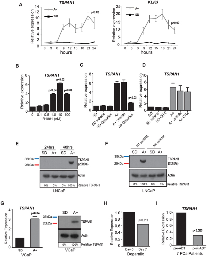
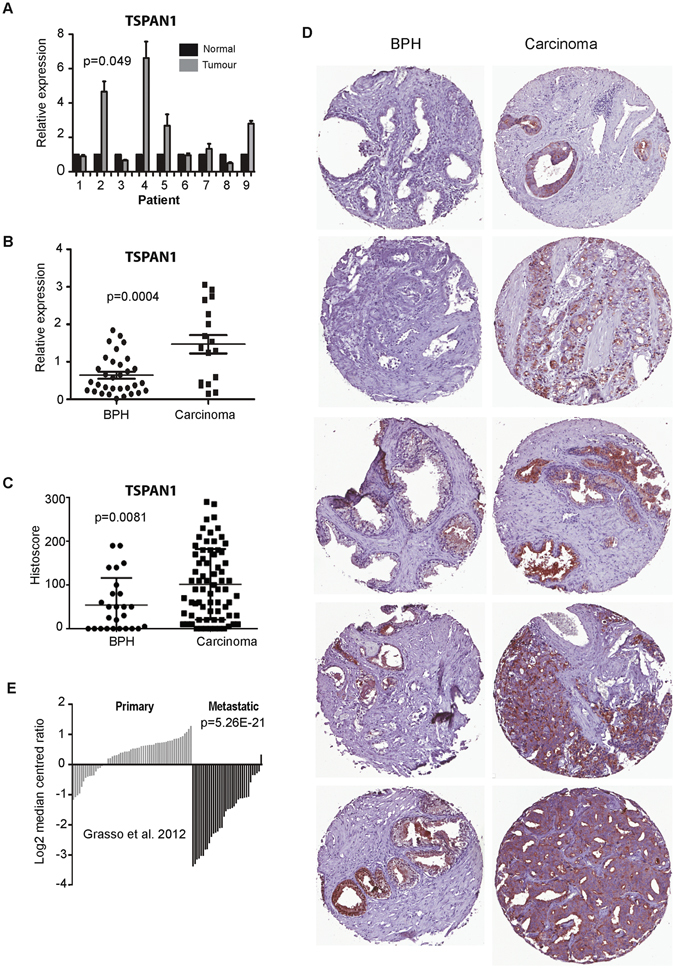
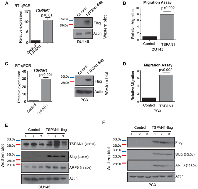
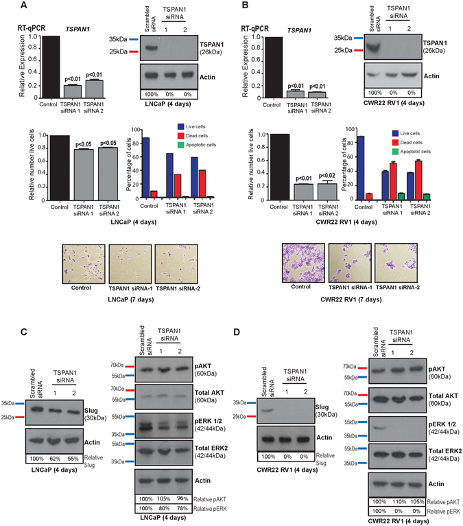
Cell migration drives cell invasion and metastatic progression in prostate cancer and is a major cause of mortality and morbidity. However the mechanisms driving cell migration in prostate cancer patients are not fully understood. We previously identified the cancer-associated cell migration protein Tetraspanin 1 (TSPAN1) as a clinically relevant androgen regulated target in prostate cancer. Here we find that TSPAN1 is acutely induced by androgens, and is significantly upregulated in prostate cancer relative to both normal prostate tissue and benign prostate hyperplasia (BPH). We also show for the first time, that TSPAN1 expression in prostate cancer cells controls the expression of key proteins involved in cell migration. Stable upregulation of TSPAN1 in both DU145 and PC3 cells significantly increased cell migration and induced the expression of the mesenchymal markers SLUG and ARF6. Our data suggest TSPAN1 is an androgen-driven contributor to cell survival and motility in prostate cancer.


Conflict of interest statement

The authors declare that they have no competing interests.

Figures

Figure 1
Figure 1
TSPAN1 is regulated by androgens in in vitro and in vivo. (A) Analysis of TSPAN1 and KLK3 (PSA) mRNA in LNCaP cells treated with androgens (A+) over a 24 hour time course. (B) Upregulation of TSPAN1 is also evident in LNCaP cells treated with 1 to 100 nM of R1881. (C) Induction of TSPAN1 mRNA by the AR is inhibited in the presence of 10 μM of the anti-androgen Casodex (bicalutamide) (lane 6). (D) The increase in TSPAN1 mRNA expression in response to androgens is still seen in the presence of 1 μg/ml cycloheximide (CHX). Relative TSPAN1 expression was detected by real-time PCR and normalised to three reference genes. (E) Expression of TSPAN1 protein is induced in LNCaP cells treated with 10 nM R1881 for 48 hours as detected by western blot. Actin was used as a loading control. (F) Depletion of AR protein in LNCaP cells by esiRNA shows that when the AR is depleted there is no induction of the TSPAN1 protein in response to androgens. (G) Real-time PCR and western blot analysis of TSPAN1 mRNA and protein in VCaP cells grown in steroid deplete (SD) or 10 nM R1881 (androgens, A+) treated conditions for 48 hours. (H) Analysis of TSPAN1 mRNA expression before and after 7 days of serum/tissue androgen depletion in prostate cancer patients using degarelix. (I) Analysis of TSPAN1 expression in previously published RNA-Seq data shows that TSPAN1 mRNA is significantly down-regulated in 7 PCa patients following androgen deprivation treatment (ADT). The Y axis shows the relative expression level of TSPAN1 (calculated by comparing the FPKM expression values of genes after ADT to the value before treatment). Please note: cropped western blots are displayed in the figure, but full length blots are included in the Supplementary Information file.
Figure 2
Figure 2
TSPAN1 is upregulated in prostate cancer tissue. (A) Real-time PCR analysis of TSPAN1 mRNA from normal and matched prostate cancer tissue from 9 patients obtained from radical prostatectomy. (B) We also analysed 32 benign samples from patients with benign prostate hyperplasia (BPH) and 17 malignant samples from transurothelial resection of the prostate (TURP) samples. (C) Analysis of TSPAN1 protein levels in patients with BPH or prostate cancer by Tissue Microarray (TMA). (D) Representative cores from each TMA. (E) TSPAN1 gene expression levels in prostate tissue from the primary or metastatic site in data published by Grasso et al..
Figure 3
Figure 3
TSPAN1 increases prostate cancer cell migration and induces the expression of Slug and ARF6. (A,C) Validation of TSPAN1 overexpression in DU145 and PC3 cells by real-time PCR and western blot. (B,D) Migration assays for DU145 and PC3 cells overexpressing TSPAN1. (E,F) Western blot analysis of Slug and ARF6 protein expression in DU145 and PC3 cells overexpressing TSPAN1. Please note: cropped western blots are displayed in the figure, but full length blots are included in the Supplementary Information file.
Figure 4
Figure 4
TSPAN1 is essential for prostate cancer cell viability. Depletion of TSPAN1 in (A) LNCaP and (B) CWR22 RV1 cells by transient transfection with siRNA. Protein depletion was carried out using two different siRNAs in cells grown in full media. 96 hours after transfection knockdown of TSPAN1 was confirmed by real-time PCR and western blot and the relative number of live cells was calculated. Representative crystal violet stained images are shown below for each cell line after 7 days (due to a dramatic decrease in cell viability we were unable to harvest enough live cells to confirm TSPAN1 knockdown after 96 hours). (C) Western blot analysis of various proteins in LNCaP and (D) CWR22 RV1 cells following siRNA mediated protein depletion of TSPAN1 for 96 hours. Please note: cropped western blots are displayed in the figure, but full length blots are included in the Supplementary Information file.

Similar articles

Cited by

References

    1. Hanahan D, Weinberg RA. The hallmarks of cancer. Cell. 2000;100:57–70. doi: 10.1016/S0092-8674(00)81683-9. - DOI - PubMed
    1. Hanahan D, Weinberg RA. Hallmarks of cancer: the next generation. Cell. 2011;144:646–674. doi: 10.1016/j.cell.2011.02.013. - DOI - PubMed
    1. Mehlen P, Puisieux A. Metastasis: a question of life or death. Nat Rev Cancer. 2006;6:449–458. doi: 10.1038/nrc1886. - DOI - PubMed
    1. Center MM, et al. International variation in prostate cancer incidence and mortality rates. Eur Urol. 2012;61:1079–1092. doi: 10.1016/j.eururo.2012.02.054. - DOI - PubMed
    1. Livermore KE, Munkley J, Elliott DJ. Androgen receptor and prostate cancer. AIMS Molecular Science. 2016;3:280–299. doi: 10.3934/molsci.2016.2.280. - DOI

Publication types

MeSH terms