Review
. 2016 Nov 24:8:23.
doi: 10.1186/s41479-016-0023-9. eCollection 2016.

Tuberculosis exposure, infection and disease in children: a systematic diagnostic approach

Affiliations
Review

Tuberculosis exposure, infection and disease in children: a systematic diagnostic approach

Claudia L Roya-Pabon et al. Pneumonia (Nathan). .

Abstract

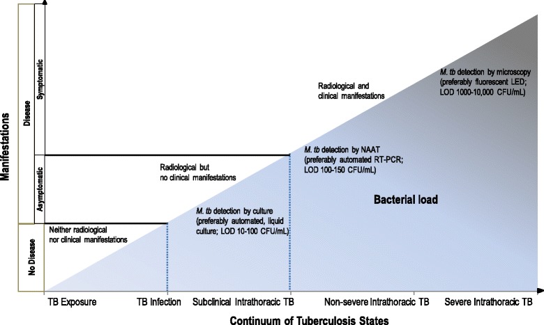
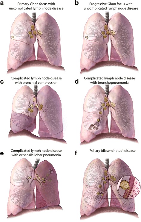
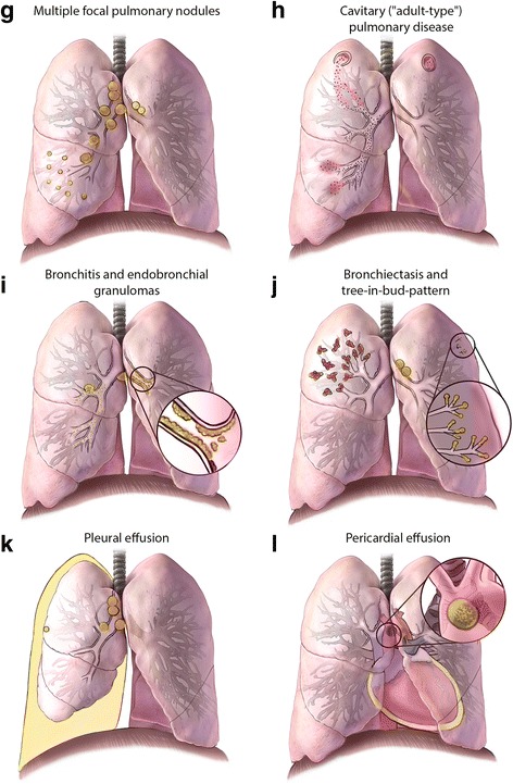
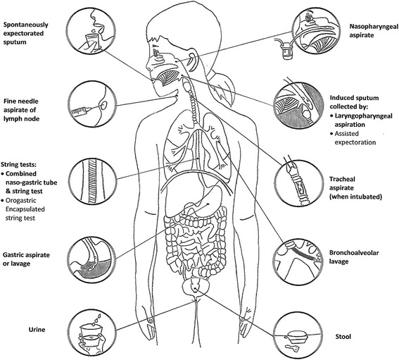
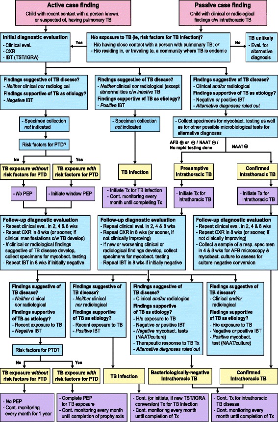
The accurate diagnosis of tuberculosis (TB) in children remains challenging. A myriad of common childhood diseases can present with similar symptoms and signs, and differentiating between exposure and infection, as well as infection and disease can be problematic. The paucibacillary nature of childhood TB complicates bacteriological confirmation and specimen collection is difficult. In most instances intrathoracic TB remains a clinical diagnosis. TB infection and disease represent a dynamic continuum from TB exposure with/without infection, to subclinical/incipient disease, to non-severe and severe disease. The clinical spectrum of intrathoracic TB in children is broad, and the classification of clinical, radiological, endoscopic, and laboratory findings into recognized clinical syndromes allows a more refined diagnostic approach in order to minimize both under- and over-diagnosis. Bacteriological confirmation can be improved significantly by collecting multiple, high-quality specimens from the most appropriate source. Mycobacterial testing should include traditional smear microscopy and culture, as well as nucleic acid amplification testing. A systematic approach to the child with recent exposure to TB, or with clinical and radiological findings compatible with this diagnosis, should allow pragmatic classification as TB exposure, infection, or disease to facilitate timely and appropriate management. It is important to also assess risk factors for TB disease progression and to undertake follow-up evaluations to monitor treatment response and ongoing evidence supporting a TB, or alternative, diagnosis.

Keywords: Algorithm; Diagnostic techniques and procedures; Latent tuberculosis; Risk factors; Specimen handling.


Figures

Fig. 1
Fig. 1
Continuum of TB states and correlations with bacterial load and with radiological and clinical manifestations. CFU: colony-forming units; LED: light-emitting diode; LOD: limit of detection; mL: milliliter; NAAT: nucleic acid amplification test; RT-PCR: real-time polymerase chain reaction. Adapted from C.M. Perez-Velez. Diagnosis of Intrathoracic Tuberculosis in Children. In: Handbook of Child and Adolescent Tuberculosis (p. 149), J.R. Starke and P.R. Donald (Eds.), 2016, New York, NY: Oxford University Press. Copyright by Oxford University Press [15]. Adapted with permission
Fig. 2
Fig. 2
Illustrations of radiological patterns caused by intrathoracic TB in children. Panel a. Primary Ghon focus with uncomplicated lymph node disease. Hilar and mediastinal lymphadenopathy associated with an ipsilateral peripheral nodule, or “Ghon focus” (right lung); these nodules are often subpleural with an overlying pleural reaction. Panel b. Progressive Ghon focus with uncomplicated lymph node disease. A Ghon focus with cavitation (right lung), which is seen almost exclusively in infants and immunocompromised children; other elements of the Ghon complex are also visible. Panel c. Complicated lymph node disease with bronchial compression. Enlarged lymph nodes compressing the airway, causing either complete obstruction with lobar collapse (right middle and lower lobes), or partial obstruction with a ball-valve effect leading to hyperinflation (left upper and lower lobes). Panel d. Complicated lymph node disease with bronchopneumonia. Necrotic lymph nodes erupting into bronchus intermedius, with endobronchial spread and patchy consolidation of the middle lobe (right lung). Panel e. Complicated lymph node disease with expansile lobar pneumonia. Necrotic lymph nodes that compress and obstruct the left upper lobe bronchus and may infiltrate a phrenic nerve, causing hemidiaphragmatic palsy (left-sided); endobronchial spread causes dense consolidation of the entire lobe (left upper lobe), with displacement of the trachea and fissures and the formation of focal cavities. Panel f. Miliary (disseminated) disease. Diffuse micronodules in both lungs, which may result from lymphohematogenous spread after recent primary infection or from infiltrating a necrotic lymph node or lung lesion into a blood vessel, leading to hematogenous spread
Fig. 2
Fig. 2
Panel g. Multiple focal pulmonary nodules. Multiple focal pulmonary nodules involving the right middle lobe with enlargement of regional lymph nodes (right lung). Panel h. Cavitary (“adult-type”) pulmonary disease. Cavity formation in both upper lobes, with endobronchial spread to the right middle lobe. Nodules or cavities in apical lung segments are typical of adult-type disease and are pathologically distinct from the other cavities shown. Panel i. Bronchitis and endobronchial granulomas. Inflammation of the mucosa of main stem bronchus with purulent secretions (left lung), and a necrotic lymph node that has eroded into the right middle lobe bronchus leading to endobronchial spread and subsequent development of endobronchial granulomas extending proximally to the bronchus intermedius and main stem bronchus, and distally to the lower lobe bronchus (right lung). These findings are best visualized by bronchoscopy. Panel j. Bronchiectasis and tree-in-bud-pattern. Bronchiectasis that extensively involves the upper lobe (right lung), and shows tree-in-bud pattern observable on CT scans -- reflecting dilated centrilobular bronchioles with mucoid impaction -- involving the upper lobe (left lung). Panel k. Pleural effusion. A pleural effusion that is usually indicative of recent primary infection, with a hypersensitivity response to tuberculoprotein leaking from a subpleural Ghon focus (often not visible) into the pleural cavity; in rare cases this effusion may also result from a chylothorax. Panel l. Pericardial effusion. A pericardial effusion that occurs when tuberculoprotein leaks from a necrotic subcarinal lymph node (shown in “close-up” window) into the pericardial space; it may also occur after hematogenous spread. Conceptualization and original sketches by C.L. Roya-Pabon, MD; finished artwork by Mesa Schumacher, MA (used with permission). Adapted from C.M. Perez-Velez. Diagnosis of Intrathoracic Tuberculosis in Children. In: Handbook of Child and Adolescent Tuberculosis (p. 154–155), J.R. Starke and P.R. Donald (Eds.), 2016, New York, NY: Oxford University Press. Copyright by Oxford University Press [15]. Adapted with permission
Fig. 3
Fig. 3
Specimens for bacteriological confirmation of intrathoracic TB in children. Adapted from C.L. Roya-Pabon. Especímenes Respiratorios para el Diagnóstico Microbiológico de las Infecciones Respiratorias. In: Neumología Pediátrica (p. 179), R. Posada-Saldarriaga (Ed.), 2016, Bogotá, Colombia: Distribuna Editorial. Copyright by Distribuna Ltda. [46]. Adapted with permission
Fig. 4
Fig. 4
Proposed diagnostic and management algorithm for a child with recent exposure to, or with clinical or radiological findings compatible with TB. AFB: acid-fast bacilli testing; Cont.: continue; c/w: compatible with; CXR: chest radiography; eval.: evaluation H/o: history of; IBT: immune-based test IGRA: interferon-gamma release assay; mycobact.: mycobacterial; NAAT: nucleic acid amplification test; PEP: post-exposure prophylaxis; PTD: progression to TB disease; TB: tuberculosis; TST: tuberculin skin test; Tx: treatment; wks: weeks

References

    1. Cruz AT, Starke JR. Clinical manifestations of tuberculosis in children. Paediatr Respir Rev. 2007;8:107–17. doi: 10.1016/j.prrv.2007.04.008. - DOI - PubMed
    1. Marais BJ, Graham SM. Childhood tuberculosis: a roadmap towards zero deaths. J Paediatr Child Health. 2016;52:258–61. doi: 10.1111/jpc.12647. - DOI - PubMed
    1. Young D, Stark J, Kirschner D. Systems biology of persistent infection: tuberculosis as a case study. Nat Rev Microbiol. 2008;6:520–8. doi: 10.1038/nrmicro1919. - DOI - PubMed
    1. Barry CE, 3rd, Boshoff HI, Dartois V, Dick T, Ehrt S, Flynn J, et al. The spectrum of latent tuberculosis: rethinking the biology and intervention strategies. Nat Rev Microbiol. 2009;7:845–55. - PMC - PubMed
    1. Young DB, Gideon HP, Wilkinson RJ. Eliminating latent tuberculosis. Trends Microbiol. 2009;17:183–8. doi: 10.1016/j.tim.2009.02.005. - DOI - PubMed

LinkOut - more resources