. 2017 Jul-Aug;23(4):272-281.
doi: 10.5152/dir.2017.16364.

Shining light in a dark landscape: MRI evaluation of unusual localization of endometriosis

Affiliations

Shining light in a dark landscape: MRI evaluation of unusual localization of endometriosis

Benedetta Gui et al. Diagn Interv Radiol. 2017 Jul-Aug.

Abstract

Endometriosis is a disease distinguished by the presence of endometrial tissue outside the uterine cavity with intralesional recurrent bleeding and resulting fibrosis. The most common locations for endometriosis are the ovaries, pelvic peritoneum, uterosacral ligaments, and torus uterinus. Typical symptoms are secondary dysmenorrhea and cyclic or chronic pelvic pain. Unusual sites of endometriosis may be associated with specific symptoms depending on the localization. Atypical pelvic endometriosis localizations can occur in the cervix, vagina, round ligaments, ureter, and nerves. Moreover, rare extrapelvic endometriosis implants can be localized in the upper abdomen, subphrenic fold, or in the abdominal wall. Magnetic resonance imaging (MRI) represents a problem-solving tool among other imaging modalities. MRI is an advantageous technique, because of its multiplanarity, high contrast resolution, and lack of ionizing radiation. Our purpose is to remind the radiologists the possibility of atypical pelvic and extrapelvic endometriosis localizations and to illustrate the specific MRI findings. Endometriotic tissue with hemorrhagic content can be distinguished from adherences and fibrosis on MRI imaging. Radiologists should keep in mind these atypical localizations in patients with suspected endometriosis, in order to achieve the diagnosis and to help the clinicians in planning a correct and complete treatment strategy.


Conflict of interest statement

Conflict of interest disclosure

The authors declared no conflicts of interest.

Figures
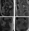
Figure 1. a–d
Figure 1. a–d
A 45-year-old woman complaining of cyclic abdominal pain and spotting (a, b) and a second case of a 40-year-old woman complaining of spotting (c, d). Sagittal T2-weighted image (a) and T1-weighted image with fat-suppression (b) show a small bright area in the cervix, within the cervical stroma, hyperintense in both sequences as hemorrhagic content (white arrow). The patient has concomitant multiple signs of endometriosis on the left ovary (b, white arrowhead). In the second case, the sagittal T2-weighted image (c) and T1-weighted image with fat-suppression (d) show a focal area in the cervical stroma that appears hyperintense on T2-weighted images and isointense on T1-weighted images as fluid continent (c, d, white arrow). In spite of the MRI appearance, cervical biopsy reveals endometriotic hyperplasia.
Figure 2. a–c
Figure 2. a–c
A 35-year-old woman with history of painful defecation, in particular during menses. Axial T2-weighted (a), sagittal T1-weighted with fat-suppression (b), and sagittal T2-weighted (c) images identify a small nodule in the posterior vaginal fornix hypointense on T2-weighted images and hyperintense on T1-weighted images due to hemorrhagic component (white arrow). Note the coexisting left intraligamentous uterine pedunculated fibroma (a, black arrow). The patient has multiple concomitant bilateral small endometriomas.
Figure 3. a, b
Figure 3. a, b
A 36-year-old patient referred with chronic pelvic pain and suspicion of pelvic endometriosis. Axial T2-weighted images (a) reveal morphologic abnormalities of intrapelvic portion of the right round ligament (white arrow) that appear thickened and shortened. Coronal T2-weighted image (b) better identifies the thickness of the distal intrapelvic portion of the right round ligament (white arrow), where it courses medial to adjacent external iliac vessels (black arrowhead) and before the inguinal canal. Anterior and posterior free pelvic fluid is also seen (white star). Note the isthmocele within the anterior wall of the uterus (a, white arrowhead). MRI examination leads the suspicion of deep endometriosis with right round ligament involvement.
Figure 4. a, b
Figure 4. a, b
A 28-year-old woman complaining of cyclic abdominal pain and painful tumefaction of the right groin, who underwent medical therapy for pelvic pain. Axial T2-weighted image (a) shows a hypointense area with spiculated margins in the right inguinal region (white arrow). The mass is strictly adjacent to the extrapelvic portion of the right round ligament of the uterus, where it passes through the inguinal canal. Axial T1-weighted image with fat-suppression (b) shows the isointense signal of the lesion (white arrow) with very small and mild hyperintense foci in the caudal portion, related to hemorrhagic components (white arrowhead). The lesion is suspicious for extrapelvic localization of deep endometriosis of the round ligament at the level of the canal of Nuck.
Figure 5. a, b
Figure 5. a, b
A 30-year-old woman complaining of cyclic pelvic pain and discomfort during urination. Coronal T2-weighted (a) image shows focal and irregular hypointense wall thickening of the upper aspect of the bladder wall (white arrow) that obliterates the vesicouterine fold and distorts the bladder wall. The detrusor muscle seems to be infiltrated and the lesion seems to project into the lumen (black arrow). Coronal T1-weighted images with fat-suppression (b) show hyperintense foci within the mass that can be attributed to hemorrhagic content (white arrowhead). Laparotomy confirmed endometriotic localization of the vesicouterine fold with bladder wall involvement.
Figure 6. a, b
Figure 6. a, b
A 31-year-old woman referred with pelvic pain, infertility, and US suspicion of endometrioma. Coronal T2-weighted image (a) shows mostly hyperintense large left ovarian mass (white arrowheads) with inhomogeneous component in declive position. The left ureter (a, black arrow) appears stretched and striped by fibrous tissue and adhesions adjacent to the endometrioma; adhesions and fibrous tissue are characterized by hypointense signal on both T2-weighted and T1-weighted images (a, b, white arrow). This tissue causes extrinsic stenosis of the ureter with associated hydronephrosis. Axial T1-weighted image with fat-suppression (b) reveals the hyperintense signal of the mass due to hemorrhagic component, chronic blood products and coagulum whitout hemorrhagic foci.
Figure 7. a, b
Figure 7. a, b
A 39-year-old patient with cyclic abdominal and right sciatic pain. Axial T2-weighted (a) and T1-weighted fat-suppression (b) images show a right nodular lesion (white arrow) located in the fat tissue between the posterior wall of acetabulus and piriform muscle, in the anatomical space where sciatic nerve is typically found. The nodular lesion shows heterogeneous hyperintense signal on T2-weighted image and hyperintensity on T1-weighted image, due to hemorrhagic content, suspicious for endometriotic localization. Free pelvic fluid is also depicted (a, star). The sciatic endometriosis was surgically confirmed.
Figure 8. a, b
Figure 8. a, b
A 45-year-old woman with history of previous cesarean sections suffering from localized abdominal pain at the site of a palpable parietal mass. Axial T2-weighted image (a) shows the presence of a hypointense spiculated mass in the subcutaneous soft tissue of abdominal wall (white arrow). The mass is strictly adjacent to the right rectus abdominis muscle (black arrow). The coronal T1-weighted images with fat-suppression (b) show slight hyperintense signal of the mass (white arrow) with some hyperintense areas due to hemorrhagic component; the coronal image reveals another small nodular lesion (white arrowhead) depicted cranially as hyperintense signal on T1-weighted image with fat-suppression. The lesions are due to abdominal wall deep endometriosis implants. Histopathology confirmed the endometriotic nature of the lesions.
Figure 9. a, b
Figure 9. a, b
A 33-year-old woman with history of pelvic pain and previous cesarean section. Axial T2-weighted image (a) shows a small nodular intramuscular lesion (white arrow) within the right rectus abdominis muscle (black arrow) with heterogeneous and predominantly hyperintense signal. Axial T1-weighed image with fat-suppression (b) shows the hyperintense signal of the lesion due to hemorrhagic foci (white arrow). Left endometrioma is also depicted (a, b, white arrowhead). Surgery confirmed the suspicion of intramuscular endometricotic focus.
Figure 10. a, b
Figure 10. a, b
A 33-year-old woman with known pelvic endometriosis referring cyclic right shoulder and abdominal pain. Coronal (a) and axial (b) T1-weighted fat-suppressed images show multiple hyperintense areas (white arrows) underneath the diaphragmatic muscular layer adjacent to the right liver lobe, in particular contiguous to the VIIth and the VIIIth segments. The patient has a concomitant voluminous right endometrioma (a, white arrowhead). The endometriotic suspicion of multiple subphrenic implants with liver capsule retraction was confirmed at abdominal surgery.
Figure 11. a–d
Figure 11. a–d
A 30-year-old patient presenting with abdominal pain, weight loss, and tension. She underwent MRI scan of abdomen with suspicion of ovarian malignancy associated with ascites previously documented by ultrasound scan of abdomen. Sagittal T2-weighted (a) and T1-weighted fat-suppressed (b) images on the pelvis show abundant ascites (star) seen as hyperintense signal on both sequences due to hematic component. MRI depicts some peritoneal implants on the left ovarian surface and on the serosal surface of the bladder dome (black arrows), better identified on T1-weighted images as small hyperintense nodules probably indicating endometriotic peritoneal locations. Note the concomitant retro-uterine endometriotic localization, with hemorrhagic component (white arrow). As collateral findings, axial T2-weighted (c) and T1-weighted fat-suppressed (d) images of the upper abdomen demonstrate bilateral pleural effusion (white arrowheads), represented more on the right side. T1-weighted image shows two small nodular implants on the right pleural surface (d, black arrowhead) with hemorrhagic features, suspicious for pleural endometriosis. Abdominal surgery confirmed the suspicion of hemorrhagic ascites with endometriotic peritoneal implants. On the basis of MRI suspicion of pleural involvement, thoracoscopy was also performed revealing multiple endometriotic nodules of right parietal pleura.

Comment in

  • Endometrioma of the sigmoid colon presenting with intestinal obstruction.
    Demir MK, Orug T, Bayık RN. Demir MK, et al. Diagn Interv Radiol. 2018 Jan-Feb;24(1):60-61. doi: 10.5152/dir.2017.17274. Diagn Interv Radiol. 2018. PMID: 29199174 Free PMC article. No abstract available.
  • Author's Reply.
    Gui B. Gui B. Diagn Interv Radiol. 2018 Jan-Feb;24(1):60-61. doi: 10.5152/dir.2017.001. Diagn Interv Radiol. 2018. PMID: 29199175 Free PMC article. No abstract available.

Similar articles

Cited by

References

    1. Novellas S, Chassang M, Bouaziz J, Delotte J, Toullalan O, Chevallier EP. Anterior pelvic endometriosis: MRI features. Abdom Imaging. 2010;35:742–749. https://doi.org/10.1007/s00261-010-9600-1. - DOI - PubMed
    1. Siegelman ES, Oliver ER. MR imaging of endometriosis: ten imaging pearls. Radiographics. 2012;32:1675–1691. https://doi.org/10.1148/rg.326125518. - DOI - PubMed
    1. Coutinho A, Jr, Bittencourt LK, Pires CE, et al. MR imaging in deep pelvic endometriosis: a pictorial essay. Radiographics. 2011;31:549–567. https://doi.org/10.1148/rg.312105144. - DOI - PubMed
    1. Woodward PF, Sohaey R, Mezzetti TP. Endometriosis: radiologic–pathologic correlation. Radiographics. 2011;21:193–216. https://doi.org/10.1148/radiographics.21.1.g01ja14193. - DOI - PubMed
    1. Bennett GL, Slywotzky CM, Cantera M, Hecht EM. Unusual manifestations and complications of endometriosis--spectrum of imaging findings: pictorial review. AJR Am J Roentgenol. 2011;194:WS34–46. https://doi.org/10.2214/AJR.07.7142. - DOI - PubMed