. 2017 Oct;6(19):10.1002/adhm.201700202.
doi: 10.1002/adhm.201700202. Epub 2017 Jul 13.

Timed Delivery of Therapy Enhances Functional Muscle Regeneration

Affiliations

Timed Delivery of Therapy Enhances Functional Muscle Regeneration

Christine A Cezar et al. Adv Healthc Mater. 2017 Oct.

Abstract

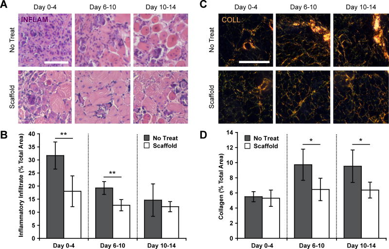
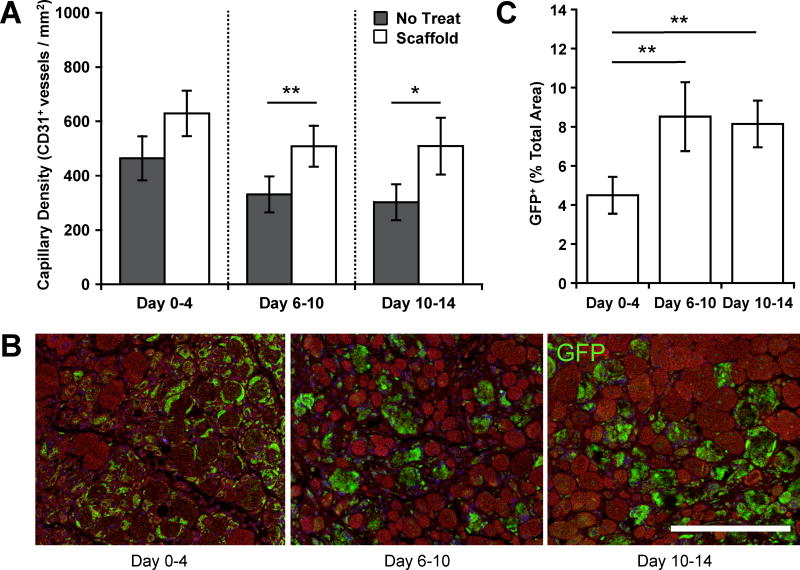
Cell transplantation is a promising therapeutic strategy for the treatment of traumatic muscle injury in humans. Previous investigations have typically focused on the identification of potent cell and growth factor treatments and optimization of spatial control over delivery. However, the optimal time point for cell transplantation remains unclear. Here, this study reports how myoblast and morphogen delivery timed to coincide with specific phases of the inflammatory response affects donor cell engraftment and the functional repair of severely injured muscle. Delivery of a biomaterial-based therapy timed with the peak of injury-induced inflammation leads to potent early and long-term regenerative benefits. Diminished inflammation and fibrosis, enhanced angiogenesis, and increased cell engraftment are seen during the acute stage following optimally timed treatment. Over the long term, treatment during peak inflammation leads to enhanced functional regeneration, as indicated by reduced chronic inflammation and fibrosis along with increased tissue perfusion and muscle contractile force. Treatments initiated immediately after injury or after inflammation had largely resolved provided more limited benefits. These results demonstrate the importance of appropriately timing the delivery of biologic therapy in the context of muscle regeneration. Biomaterial-based timed delivery can likely be applied to other tissues and is of potential wide utility in regenerative medicine.

Keywords: cell therapy; controlled delivery; ferrogel scaffolds; inflammation kinetics; magnetic biomaterials.


Figures

Figure 1
Figure 1
Severe muscle injury model results in acute inflammation that largely resolves by day 10. (A) and (C) Representative images and quantification of inflammation probe dye (DYE) accumulating at injury site at varying time points following induction of ischemia, as measured by IVIS. Regions of interest used for quantification are marked with blue ovals. Ratios of dye accumulation in injured hindlimbs to uninjured contralateral hindlimbs were calculated to account for variability associated with dye leakage from and around the sinus following retro-orbital injections.[54] Footpads of injured hindlimbs were not used for quantification due to minimal blood flow and dye accumulation downstream of ischemic injuries. (B) and (D) Representative images and quantification of inflammatory infiltrate (INFLAM) in histological cross-sections of tibialis anterior muscles stained with hematoxylin and eosin at varying time points following ischemia injury. Scale bar represents 200 µm. (E) Experimental design showing injury, scaffold implantation, and sacrifice time points. The time points at which scaffolds were stimulated to induce release of cells and factors are also indicated. Values represent the mean and standard deviation (n = 5–7). Data in C and D were compared using ANOVA with Bonferroni's post-hoc test (*p < 0.05).
Figure 2
Figure 2
Timing of therapy modulates acute inflammation and fibrosis. (A) and (B) Representative images and quantification of inflammatory infiltrate (INFLAM) in histological cross-sections of tibialis anterior muscles stained with hematoxylin and eosin. Muscles were treated at each time point via scaffold delivery (Scaffold) or left untreated (No Treat). Treatments included placement of the scaffold immediately after induction of ischemia (Day 0–4), 6 days after induction of ischemia (Day 6–10), or 10 days after induction of ischemia (Day 10–14) followed by stimulation for four consecutive days. Analysis was always performed 4 days after initiation of each treatment. (C) and (D) Representative images and quantification of collagen deposition, as assessed from picrosirius red stained cross-sections, with the same treatments and time of analysis. All scale bars represent 200 µm. Values represent the mean and standard deviation (n = 5). Data in B and Figure S4B, as well as data in D and Figure S4D were compared using ANOVA with Bonferroni's post-hoc test (*p < 0.05, **p < 0.01).
Figure 3
Figure 3
Timing of therapy modulates early angiogenesis and transplanted cell engraftment efficiency. (A) Muscle capillary densities, as assessed by CD31+ staining of tissue samples 4 days after initiation of scaffold treatment. (B) and (C) Representative images and quantification of the percentage of donor GFP myoblasts and newly formed GFP myofibers 4 days after initiation of scaffold treatment in histological cross-sections of tibialis anterior muscles. GFP donor cells remaining as single cells and GFP myofibers were quantified together. Scale bar represents 100 µm. Values represent the mean and standard deviation (n = 5). Data in A and Figure S5A, as well as data in C and Figure S5C were compared using ANOVA with Bonferroni's post-hoc test (*p < 0.05, **p < 0.01).
Figure 4
Figure 4
Long-term functional muscle regeneration results from appropriate timing of therapy. (A) and (C) Representative images and quantification of inflammatory infiltrate in histological cross-sections of tibialis anterior muscles stained with hematoxylin and eosin 6 weeks post-injury. (B) and (D) Representative images and quantification of collagen deposition from picrosirius red stained cross-sections 6 weeks post-injury. (E) Perfusion of injured hindlimbs, normalized to uninjured contralateral controls, as measured by Laser doppler perfusion imaging (LDPI). (F) Maximum contractile force following tetanic stimulation, 6 weeks post-injury of intact muscles receiving no treatment or scaffold treatment at day 0, 6, or 10. All scale bars represent 500 µm. Values represent the mean and standard deviation (n = 6). Data in C–F were compared using ANOVA with Bonferroni's post-hoc test (*p < 0.05, **p < 0.01, ***p < 0.001).

References

    1. Tedesco FS, Cossu G. Curr. Opin. Neurol. 2012;25:597. - PubMed
    1. Palmieri B, Tremblay JP, Daniele L. Pediatr. Transplant. 2010;14:813. - PubMed
    1. Smythe GM, Hodgetts SI, Grounds MD. Mol. Ther. 2000;1:304. - PubMed
    1. Fan Y, Maley M, Beilharz M, Grounds M. Muscle Nerve. 1996;19:853. - PubMed
    1. Boldrin L, Neal A, Zammit PS, Muntoni F, Morgan JE. Stem Cells. 2012;30:1971. - PMC - PubMed

MeSH terms

Substances