. 2017 Jul 13;8(7):e2927.
doi: 10.1038/cddis.2017.317.

Inhibition of autophagy as a treatment strategy for p53 wild-type acute myeloid leukemia

Affiliations

Inhibition of autophagy as a treatment strategy for p53 wild-type acute myeloid leukemia

Hendrik Folkerts et al. Cell Death Dis. .

Abstract

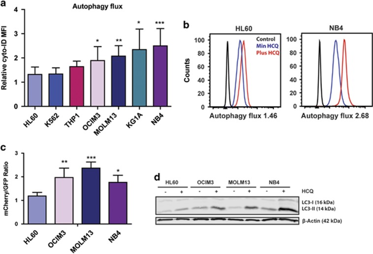
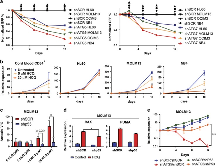
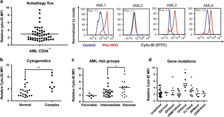
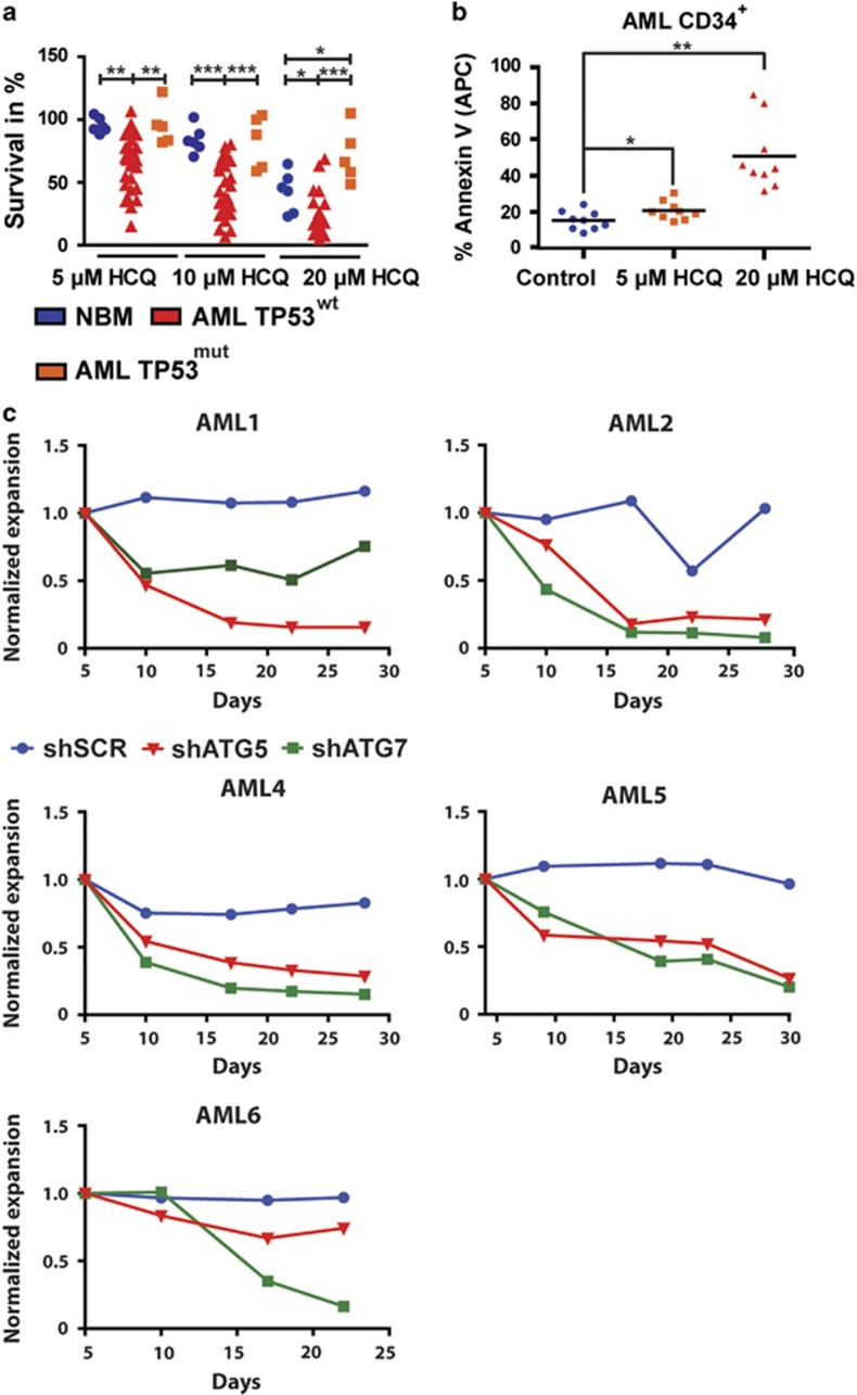
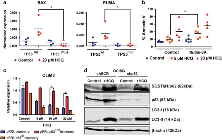
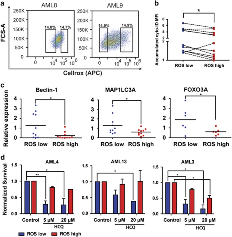
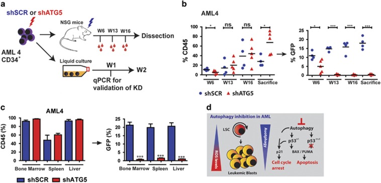
Here we have explored whether inhibition of autophagy can be used as a treatment strategy for acute myeloid leukemia (AML). Steady-state autophagy was measured in leukemic cell lines and primary human CD34+ AML cells with a large variability in basal autophagy between AMLs observed. The autophagy flux was higher in AMLs classified as poor risk, which are frequently associated with TP53 mutations (TP53mut), compared with favorable- and intermediate-risk AMLs. In addition, the higher flux was associated with a higher expression level of several autophagy genes, but was not affected by alterations in p53 expression by knocking down p53 or overexpression of wild-type p53 or p53R273H. AML CD34+ cells were more sensitive to the autophagy inhibitor hydroxychloroquine (HCQ) than normal bone marrow CD34+ cells. Similar, inhibition of autophagy by knockdown of ATG5 or ATG7 triggered apoptosis, which coincided with increased expression of p53. In contrast to wild-type p53 AML (TP53wt), HCQ treatment did not trigger a BAX and PUMA-dependent apoptotic response in AMLs harboring TP53mut. To further characterize autophagy in the leukemic stem cell-enriched cell fraction AML CD34+ cells were separated into ROSlow and ROShigh subfractions. The immature AML CD34+-enriched ROSlow cells maintained higher basal autophagy and showed reduced survival upon HCQ treatment compared with ROShigh cells. Finally, knockdown of ATG5 inhibits in vivo maintenance of AML CD34+ cells in NSG mice. These results indicate that targeting autophagy might provide new therapeutic options for treatment of AML since it affects the immature AML subfraction.


Conflict of interest statement

The authors declare no conflict of interest.

Figures

Figure 1
Figure 1
Variation in autophagy flux between different leukemic cell lines. (a) Relative accumulation of autophagosomes after overnight treatment with 20 μM HCQ measured by staining with Cyto-ID in a panel of leukemic cell lines (N=7). (b) Representative FACS plots showing mean fluorescent intensity of Cyto-ID, with or without HCQ treatment. (c) mCherry/GFP ratio in a panel of leukemic cell lines transduced with mCherry-GFP-LC3. (d) Representative western blot of LC3-II accumulation after HCQ in cell lines, β-actin was used as loading control. Error bars represent S.D.; *, ** or *** represents P<0.05, P<0.01 or P<0.001, respectively
Figure 2
Figure 2
Sensitivity for inhibition of autophagy in leukemic cells. (a) Normalized GFP percentages in leukemic cell lines transduced with shSCR-GFP, shATG5-GFP or shATG7-GFP and cultured for 12 days. (b) Cumulative growth of leukemia cell lines and cord blood-derived CD34+ cells cultured for 10 days in the presence of 0, 5 or 20 μM HCQ. (c) Percentage of Annexin-V-positive cells in shSCR or shp53 transduced MOLM13 cells, at day 4 after treatment with different concentrations of HCQ. (d) Quantitative RT-PCR for BAX and PUMA in shSCR and shP53 transduced MOLM13 cells, treated with 20 μM HCQ for 4 days. (e) Cell expansion in time of MOLM13 cells double transduced with shp53-GFP or shSCR-GFP in combination with shSCR-mCherry or shATG5-mCherry. The transduced cells were cultured for 12 days. Error bars represent S.D.; *, ** or *** represents P<0.05, P<0.01 or P<0.001, respectively
Figure 3
Figure 3
Variation in autophagy levels between different AMLs, independent of the differentiation status. (a) Left panel, for autophagic flux measurements (relative Cyto-ID accumulation) in AML CD34+ blasts (n=51), AML CD34+ cells were cultured for 3 days on MS5 stromal layer before overnight incubation with 20 μM HCQ. Right panel, representative FACS plot showing the accumulation of Cyto-ID after treatment with HCQ. (b) Autophagy flux in AMLs with normal karyotype versus complex cytogenetic abnormalities. (c) Autophagy flux in AMLs according to the various ELN risk groups. (d) Autophagy flux in AML CD34+ cells according to commonly mutated genes in AML. Error bars represent S.D.; *, ** or *** represents P<0.05, P<0.01 or P<0.001, respectively
Figure 4
Figure 4
Inhibition of autophagy triggers apoptosis in primary AML CD34+ cells. (a) Survival of normal bone marrow (NBM) CD34+, TP53wt AML CD34+ or TP53mut AML CD34+ cells were cultured for 3 days on an MS5 stromal layer before treated with 5, 10 or 20 μM HCQ for 48 h. (b) Quantification of Annexin-V percentages in AML (n=9) after treatment with 5 or 20 μM HCQ. (c) Normalized expansion of AML CD34+ cells transduced with shSCR, shATG5 or shATG7, cultured on an MS5 stromal layer. Error bars represent S.D.; *, ** or *** represents P<0.05, P<0.01 or P<0.001, respectively
Figure 5
Figure 5
TP53 mutant AMLs are resistant for HCQ-induced apoptosis. (a) Gene expression of BAX and PUMA determined by quantative RT-PCR in TP53wt (n=4) or TP53mut (n=4) AMLs. AML CD34+ cells were cultured for 3 days on an MS5 stromal layer before 72 h incubation with 20 μM HCQ. (b) Percentage of Annexin-V-positive cells in TP53wt AML CD34+ cells treated with 5 or 20 μM HCQ in conjunction with or without Nutlin-3A. (c) Cell counts of OCIM3 cells transduced with pRRL-mBlueberry, pRRL-P53mut-mBlueberry or pRRL-P53wt-mBlueberry, treated with different concentrations of HCQ. (d) Western blot showing LC3-II, sqstm1/p62 and p53 protein expression in OCIM3 cells transduced with shSCR or shP53 treated overnight with or without 20 μM HCQ. Error bars represent S.D.; * represents P<0.05
Figure 6
Figure 6
Autophagy is higher in the ROSlow population of AML blasts. (a) Representative FACS plots showing CellROX staining in freshly isolated AML CD34+ cells. (b) Relative Cyto-ID levels in ROShigh and ROSlow fractions of AML CD34+ cells (n=11). (c) Gene expression of Beclin-1 and MAP1LC3A in freshly sorted AML CD34+ROShigh and CD34+ROSlow cells. (d) Survival of FACS-sorted ROShigh and ROSlow AML CD34+ cells, cultured for 3 days on an MS5 stromal layer before treated for 48 h with different concentrations HCQ. Error bars represent S.D., * or ** represents P<0.05 or P<0.01 respectively
Figure 7
Figure 7
Knockdown of ATG5 in AML CD34+ blasts results in impaired engraftment. (a) Experimental set-up. (b) Left panel: engraftment levels measured by huCD45%. Right panel: the GFP% within huCD45+ population. Each dot represents data from a single mouse, shSCR (N=4) and shATG5 (N=5). (c) Engraftment (percentage huCD45) at time of killing in bone marrow, spleen and liver and the GFP% within the huCD45+ population. (d) Summarizing Model: LSCs are enriched in the ROSlow fraction of AML blasts. ROSlow cells maintain a higher basal autophagy flux and have a lower mitochondrial mass compared with ROShigh cells. Right part: short-term genetic or pharmaceutical Inhibition of autophagy triggered a p53-dependent apoptotic response in p53 wild-type AMLs, which was severely dampened in p53 mutant AMLs. Error bars represent S.D.; * or ***represents P<0.05 or P<0.001, respectively

References

    1. Shen Y, Zhu Y-M, Fan X, Shi JY, Wang QR, Yan XJ et al. Gene mutation patterns and their prognostic impact in a cohort of 1185 patients with acute myeloid leukemia. Blood 2011; 118: 5593–5603. - PubMed
    1. Wouters BJ, Delwel R. Epigenetics and approaches to targeted epigenetic therapy in acute myeloid leukemia. Blood 2016; 127: 42–52. - PubMed
    1. Rose D, Haferlach T, Schnittger S, Takahashi N, Yamashita T. Subtype-specific patterns of molecular mutations in acute myeloid leukemia. Leukemia 2017; 31: 11–17. - PubMed
    1. Testa U, Riccioni R. Deregulation of apoptosis in acute myeloid leukemia. Haematologica 2007; 92: 81–94. - PubMed
    1. Bosman MCJ, Schepers H, Jaques J, Brouwers-Vos AZ, Quax WJ, Schuringa JJ et al. The TAK1-NF-κB axis as therapeutic target for AML. Blood 2014; 124: 3130–3140. - PubMed

Publication types

MeSH terms