. 2017 Aug 30;139(34):11734-11744.
doi: 10.1021/jacs.7b01283. Epub 2017 Aug 21.

Structural Insights into Thioether Bond Formation in the Biosynthesis of Sactipeptides

Affiliations

Structural Insights into Thioether Bond Formation in the Biosynthesis of Sactipeptides

Tyler L Grove et al. J Am Chem Soc. .

Abstract

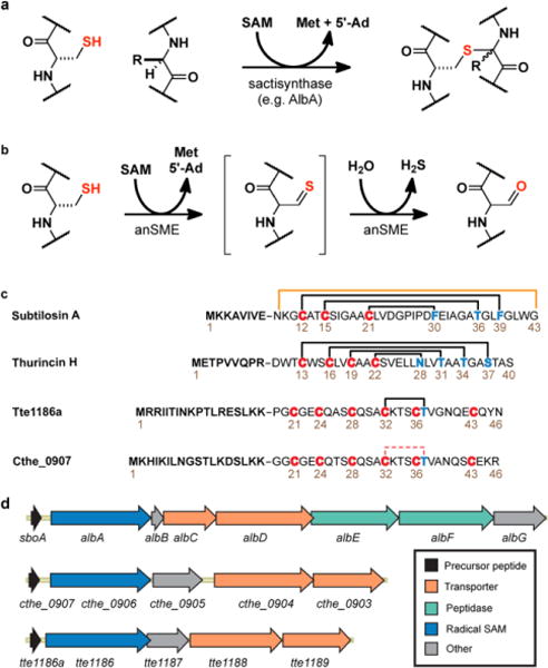
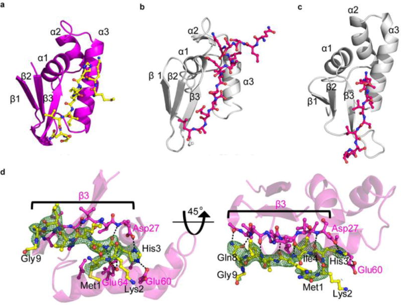
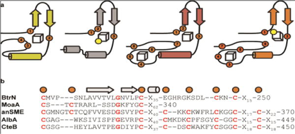
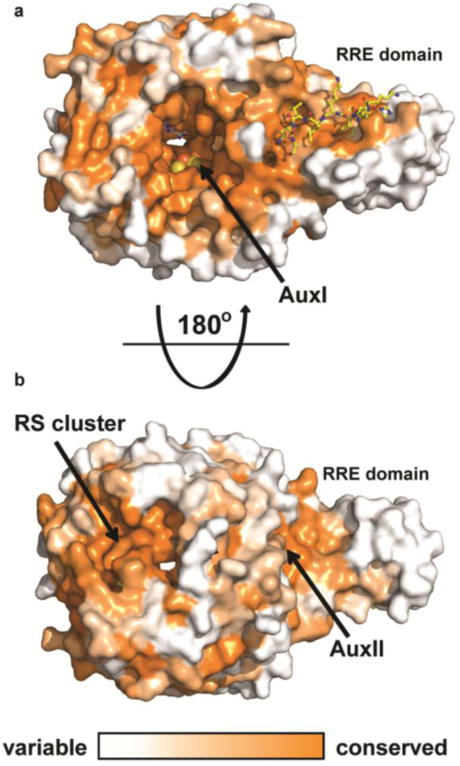
Sactipeptides are ribosomally synthesized peptides that contain a characteristic thioether bridge (sactionine bond) that is installed posttranslationally and is absolutely required for their antibiotic activity. Sactipeptide biosynthesis requires a unique family of radical SAM enzymes, which contain multiple [4Fe-4S] clusters, to form the requisite thioether bridge between a cysteine and the α-carbon of an opposing amino acid through radical-based chemistry. Here we present the structure of the sactionine bond-forming enzyme CteB, from Clostridium thermocellum ATCC 27405, with both SAM and an N-terminal fragment of its peptidyl-substrate at 2.04 Å resolution. CteB has the (β/α)6-TIM barrel fold that is characteristic of radical SAM enzymes, as well as a C-terminal SPASM domain that contains two auxiliary [4Fe-4S] clusters. Importantly, one [4Fe-4S] cluster in the SPASM domain exhibits an open coordination site in absence of peptide substrate, which is coordinated by a peptidyl-cysteine residue in the bound state. The crystal structure of CteB also reveals an accessory N-terminal domain that has high structural similarity to a recently discovered motif present in several enzymes that act on ribosomally synthesized and post-translationally modified peptides (RiPPs), known as a RiPP precursor peptide recognition element (RRE). This crystal structure is the first of a sactionine bond forming enzyme and sheds light on structures and mechanisms of other members of this class such as AlbA or ThnB.


Conflict of interest statement

Notes

The authors declare no competing financial interest.

Figures

Figure 1
Figure 1
Introduction to sactipeptides. a) Formation of sactionine thioether linkages found in sactipeptides. b) Formation of formyl glycine from cysteine by anSME c) Comparison of known sactipeptides to the bridge formed in CteA. d) Gene clusters of some known sactipeptide producers.
Figure 2
Figure 2
MS analysis of CteA modified by CteB. a) Expected masses of CteA modified by CteB and alkylated with NEM. b) MS of CteA modified by CteB. In red is 1 eq. of CteA treated with 1 eq. of CteB while in black is CteA unmodified. The difference between corresponding charge states is that of one sactionine bridge or two hydrogen atoms. c) MS of CteA product alkylated with NEM after first being modified by CteB. In red is 1 eq. of CteA treated with 1 eq. of CteB then NEM, while in black is CteA treated with just NEM. The difference between corresponding charge states is that of one sactionine bridge and one NEM modification. See Table S3 for expected exact masses. d) MS/MS analysis (+7 charge state) of where the sactionine bridge is forming in modified CteA.
Figure 3
Figure 3
Structure of CteB. a) Overall structure of CteB. The β66 core of the RS domain (green) contains one [4Fe-4S] cluster that coordinates one molecule of SAM. The C-terminal SPASM domain (orange) contains the [4Fe-4S] clusters Aux I and Aux II and comprises residues 344 - 432. The N-terminal RRE domain (magenta) of CteB provides the binding specificity for the peptide substrate leader sequence of CteA (yellow, stick representation). b) Topology figure of CteB. c) Zoom of [4Fe-4S] clusters present in CteB along with their distances from one another. The distance from RS to Aux I is 14.4 Å while the distance from Aux I to Aux II is 11.6Å. RS, radical SAM cluster, Aux I, and, Aux II d) The Fo-Fc omit map contoured to 3.0 σ (green mesh) of Gly20 and Cys21 from CteA-M1-C21 substrate bound to Aux I. The distance between the Fe and Sϒ of Cys21 is 2.7 Å. The 2Fo-Fc map (blue mesh) for the Aux I cluster is contoured to 2.0 σ.
Figure 4
Figure 4
Leader peptide and binding to RRE of CteB. Comparison of RRE domains from CteB (a), LynD (b), NisB (c). d) Simulated annealing omit composite map (2Fo-Fc) contoured to 1.0 σ of residues 1 – 9 of the leader peptide (yellow sticks) of CteA. Residues from CteA involved in binding of the leader peptide are shown in yellow. Hydrogen bond interactions are shown as dashed lines. β3 from the RRE domain is shown in pink sticks. β3 forms extensive hydrogen bonds to the leader peptide of CteA, while α3 forms mostly hydrophobic interactions, with the exception of Glu60 and Glu64. For full list of interactions and distances see supplementary Table S6.
Figure 5
Figure 5
Comparison of Aux I and Aux II clusters. a) Topology diagrams of known crystallized enzymes that hold either one or both Aux I and Aux II clusters. Yellow-BtrN, gray-MoaA, red-anSME, and orange-CteB. b) Sequence alignments of those domains.
Figure 6
Figure 6
Proposed mechanisms of sactionine bridge formation. a) Mechanisms describing either separate activation of the bridging partner α-carbon and the cysteine sulfur by distinct [4Fe-4S] clusters followed by attack of the carbon centered radical on the coordinated sulfur atom (Mechanism A) or the intermediate α-carbon radical undergoes a one-electron oxidation to the ketoimine which is then subject to nucleophilic attack of the cysteine sulfur (Mechanism B). b) Proposed binding of substrates in their enzymes. c) Rosetta model of CteA:CteB complex with Cys32 ligated to Aux I. c) Computational model generated with Rosetta showing possible interactions between CteA (yellow) and CteB. In the model Cys32 from CteA ligates the free coordination site on Aux I and Thr37 is placed in close proximity to where the 5’-dA radical is formed from SAM (gray)
Figure 7
Figure 7
Conservation of CteB homologs. Surface map (ConSURF server) of sequence conservation based on 150 sequences with homology ranging from 35% to 90% identity. Conservation scores are based on Bayesian method. a) The highest sequence conservation can be found around the active site and peptide binding surface of the RRE domain. b) 180 ° rotation showing the bottom of CteB. A patch of highly conserved residues is found around the RS and Aux II clusters. These sites may have a role in the recognition of redox partners.

References

    1. Arnison PG, Bibb MJ, Bierbaum G, Bowers AA, Bugni TS, Bulaj G, Camarero JA, Campopiano DJ, Challis GL, Clardy J, Cotter PD, Craik DJ, Dawson M, Dittmann E, Donadio S, Dorrestein PC, Entian K-D, Fischbach MA, Garavelli JS, Göransson U, Gruber CW, Haft DH, Hemscheidt TK, Hertweck C, Hill C, Horswill AR, Jaspars M, Kelly WL, Klinman JP, Kuipers OP, Link AJ, Liu W, Marahiel MA, Mitchell DA, Moll GN, Moore BS, Muller R, Nair SK, Nes IF, Norris GE, Olivera BM, Onaka H, Patchett ML, Piel J, Reaney MJT, Rebuffat S, Ross RP, Sahl H-G, Schmidt EW, Selsted ME, Severinov K, Shen B, Sivonen K, Smith L, Stein T, Süssmuth RD, Tagg JR, Tang G-L, Truman AW, Vederas JC, Walsh CT, Walton JD, Wenzel SC, Willey JM, van der Donk WA. Nat Prod Rep. 2012;30(1):108. - PMC - PubMed
    1. Ortega MA, van der Donk WA. Cell Chem Biol. 2016;23(1):31. - PMC - PubMed
    1. Dunbar KL, Mitchell DA. ACS Chem Biol. 2013;8(3):473. - PMC - PubMed
    1. McIntosh JA, Donia MS, Schmidt EW. Nat Prod Rep. 2009;26(4):537. - PMC - PubMed
    1. Bowers AA, Acker MG, Koglin A, Walsh CT. J Am Chem Soc. 2010;132(21):7519. - PMC - PubMed

Publication types

MeSH terms