. 2017 Jul 12;22(1):120-128.e4.
doi: 10.1016/j.chom.2017.06.014.

Diabetes Enhances IL-17 Expression and Alters the Oral Microbiome to Increase Its Pathogenicity

Affiliations

Diabetes Enhances IL-17 Expression and Alters the Oral Microbiome to Increase Its Pathogenicity

E Xiao et al. Cell Host Microbe. .

Abstract

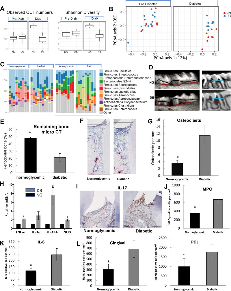
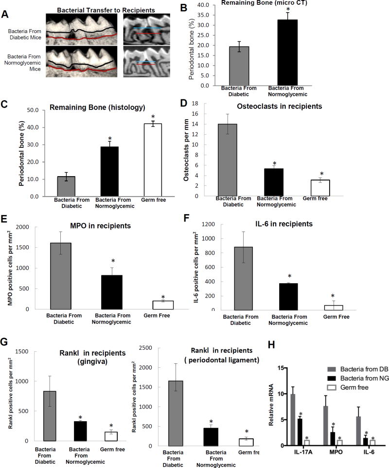
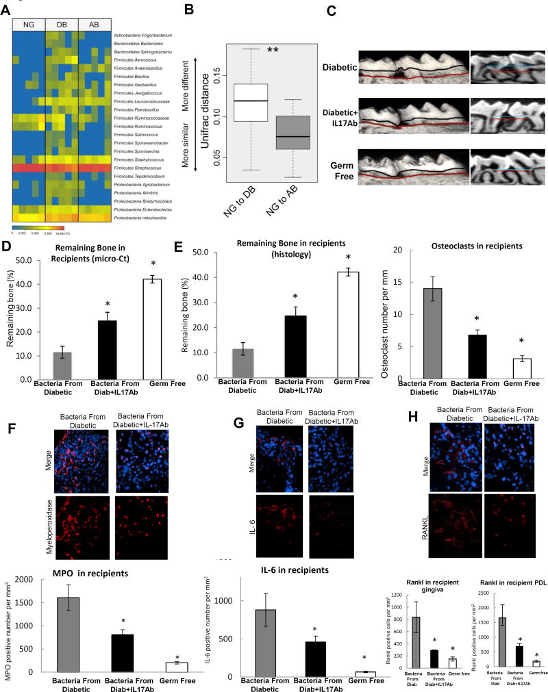
Diabetes is a risk factor for periodontitis, an inflammatory bone disorder and the greatest cause of tooth loss in adults. Diabetes has a significant impact on the gut microbiota; however, studies in the oral cavity have been inconclusive. By 16S rRNA sequencing, we show here that diabetes causes a shift in oral bacterial composition and, by transfer to germ-free mice, that the oral microbiota of diabetic mice is more pathogenic. Furthermore, treatment with IL-17 antibody decreases the pathogenicity of the oral microbiota in diabetic mice; when transferred to recipient germ-free mice, oral microbiota from IL-17-treated donors induced reduced neutrophil recruitment, reduced IL-6 and RANKL, and less bone resorption. Thus, diabetes-enhanced IL-17 alters the oral microbiota and renders it more pathogenic. Our findings provide a mechanistic basis to better understand how diabetes can increase the risk and severity of tooth loss.

Keywords: IL-17; bone loss; diabetes; dysbiosis; microbiota; osteoclast; pathogen; periodontitis.


Figures

Figure 1
Figure 1. Diabetes alters the composition of the oral microbiome and increases periodontal inflammation and bone loss
Db/db type 2 diabetes prone and lean normoglycemic control littermates (db/+) were examined. A: Alpha diversity was assessed in the microbiota of 9 normoglycemic and 10 diabetes-prone mice before hyperglycemia and 8 normoglycemic and 9 diabetic mice after the latter developed hyperglycemia. B: Beta (Unweighted Unifrac) diversity was assessed in 9 normoglycemic and 10 diabetes-prone mice before and 8 normoglycemic and 9 diabetic mice after the development of hyperglycemia. C: Prominent bacterial taxa in 9 normoglycemic and 10 diabetes-prone mice before and 8 normoglycemic and 9 diabetic mice after the development of hyperglycemia. D: Micro-CT 3D reconstructions and sagittal slice views of normoglycemic and diabetic mice. The distance between horizontal lines represents the distance between a fixed landmark on tooth surface (cemental-enamel junction) and bone crest. E: Percent periodontal bone remaining in normoglycemic and diabetic mice measured by micro-CT. A minimum of six maxillary samples from different mice were examined per group with the analysis performed 3 times with similar results. F: TRAP stained histologic sections of periodontal tissue from diabetic and normoglycemic mice. The horizontal line represents the position of cemental enamel junction and the vertical line extends from cemental enamel junction to bone height. Osteoclasts are stained red. G: Osteoclast numbers in TRAP stained histologic sections per mm bone length. Two slides from a minimum of 6 maxillary samples were analyzed per group from different animals and the analysis was carried out twice with similar results. H: mRNA was extracted from the gingiva of molar teeth, RNA extracted and mRNA levels of TNF, IL-1α, IL-17A and iNOS were measured by real-time PCR in normoglycemic (NG) and diabetic (DB) groups. A minimum of six maxillary gingival tissue samples per group were examined from different mice by real-time PCR, which was performed three times with similar results. I: Representative immunostaining of histologic sections of normoglycemic and diabetic mice with anti-IL-17A antibody. Horizontal line = 100 µm. J: Immunofluorescence with antibody to myeloperoxidase. Myeloperoxidase-positive cells were counted in gingival connective tissue. K: Immunofluorescence with IL-6 antibody. IL-6 immunopositive cells were counted in gingival connective tissue. L: Immunofluorescence with antibody specific for RANKL. RANKL immunopositive cells were counted in the gingival connective tissue and periodontal ligament. Nuclei were detected with DAPI counterstain. Data are expressed as number of positive cells per mm2 gingival connective tissue or periodontal ligament. For immunofluorescence analysis (I–K) two to three slides were analyzed from at least eight maxillary samples per group, each from a different animal. Immunofluorescence with each control antibody was negative (not shown). Original magnification of fluorescent images 400×. * indicates statistical difference (p<0.05) between diabetic and matched normoglycemic mice.
Figure 2
Figure 2. Diabetes increases the pathogenicity of bacteria transferred to normal germ-free mice
Bacteria were collected by swabbing the teeth and adjacent periodontal tissue of 5 diabetic (db/db) mice or 5 normoglycemic controls (db/+). Collected bacteria were transferred twice with one day in between to normal germ-free mice that had ligatures placed between the left maxillary 1st and 2nd molars. Mice were euthanized and periodontal tissues from recipient normal germ-free mice were examined one week after the first bacterial transfer. A: Representative images of 3D micro-CT reconstructions of periodontal bone following bacterial transfer. B: Periodontal bone was measured by micro-CT following bacterial transfer. Five maxillary samples from 5 different mice per group were analyzed and the analysis was performed 3 times. C: Representative histologic sections of germ-free mice that received bacteria from normoglycemic and diabetic donor mice were examined by TRAP staining. D: The number of TRAP-stained bone-lining multinucleated osteoclasts was counted per mm bone length. E: Immunofluorescence with an antibody specific for myeloperoxidase to quantify neutrophils per mm2 gingival connective tissue after bacterial transfer to normal germ-free mice. Immunofluorescence with control antibody was negative. F: Immunofluorescence with anti-IL-6 antibody following transfer of bacteria to normal germ-free mice. Positive cells were counted per mm2 gingival connective tissue or periodontal ligament. G: Immunofluorescence with antibody specific for RANKL. RANKL immunopositive cells were counted in the gingival connective tissue and in the periodontal ligament. Immunofluorescence with control antibody was negative. Original magnification of fluorescent images 400×. H. mRNA levels in gingiva of recipient mice after transfer of bacteria from diabetic (DB) mice or normoglycemic mice (NG) or germ-free mice without bacterial transfer. Five gingival samples from different mice were examined per group and real-time PCR was performed. The results are representative of three analyses. For histomorphometric analysis (C & D) two to three slides were examined per maxilla with maxillary samples obtained from different animals. Sections were examined twice with similar results. For immunofluorescence studies (E–G) two to three slides were examined per maxilla from five different mice per group and the analysis was carried out twice with similar results. Nuclei were detected by DAPI counterstain. * indicates significantly different from recipients that received bacteria from diabetic mice (p<0.05).
Figure 3
Figure 3. Local injection of IL-17 antibody reduces microbial pathogenicity induced by diabetes
A & B: The oral bacterial composition from normoglycemic (NG), diabetic (DB) and diabetic IL-17A antibody treated mice (AB) was examined. A. Heat map of oral bacteria from 5 NG, 5 DB and 5 AB groups. B: Unifrac distance of oral bacterial communities from NG, DB and AB groups. C–H: Bacteria from db/db diabetic mice treated with local injection of IL-17A antibody or matched control antibody were transferred to normal germ-free mice that had ligatures placed between the left maxillary 1st and 2nd molars. Mice were euthanized 1 week after bacterial transfer and periodontal tissues were examined. C: Three-dimensional micro-CT reconstruction and sagittal images. D: Periodontal bone remaining after bacterial transfer to normal germ-free mice. Five maxillary samples were examined from 5 different mice per group and the analysis was performed 3 times with similar results. E: Histologic sections were examined to quantify the interdental bone between the first and second molars. Bone lining multi-nucleated TRAP stained osteoclasts were counted per mm bone length. For each measurement two slides were examined from five maxillary samples per group and each maxilla was obtained from a different mouse. The results are representative of two separate analyses. F: Immunofluorescence with anti-myeloperoxidase antibody to quantify neutrophils per mm2 gingival connective tissue following the transfer of bacteria to normal germ-free mice. G: Immunofluorescence with anti-IL-6 antibody. H: Immunofluorescence with antibody specific to RANKL. RANKL positive cells were counted per mm2 in gingival connective tissue or periodontal ligament. For immunofluorescence (F–H) two to three slides were examined from 5 maxillary samples, each from a different mouse. The analysis was carried out twice with similar results. Nuclei were detected by DAPI counterstain. Immunofluorescence with each control antibody was negative (not shown). * Statistical difference (p<0.05) when compared to germ-free normoglycemic mice that received bacteria from diabetic donor mice. Original magnification of fluorescent images 400×.

Similar articles

Cited by

References

    1. Abusleme L, Dupuy AK, Dutzan N, Silva N, Burleson JA, Strausbaugh LD, Gamonal J, Diaz PI. The subgingival microbiome in health and periodontitis and its relationship with community biomass and inflammation. The ISME journal. 2013;7:1016–1025. - PMC - PubMed
    1. Abusleme L, Moutsopoulos NM. IL-17; overview and role in oral immunity and microbiome. Oral Dis. 2016;20:12598. - PMC - PubMed
    1. Anderson M. A new method for non-parametric multivariate analysis of variance. Austral Ecology. 2001:32–46.
    1. Andriankaja OM, Galicia J, Dong G, Xiao W, Alawi F, Graves DT. Gene Expression Dynamics during Diabetic Periodontitis. Journal of dental research. 2012;91:1160–1165. - PMC - PubMed
    1. Camelo-Castillo A, Novoa L, Balsa-Castro C, Blanco J, Mira A, Tomas I. Relationship between periodontitis-associated subgingival microbiota and clinical inflammation by 16S pyrosequencing. J Clin Periodontol. 2015;42:1074–1082. - PubMed

MeSH terms

LinkOut - more resources