

The genetics of hair-cell function in zebrafish
- PMID: 28705044
- PMCID: PMC6080859
- DOI: 10.1080/01677063.2017.1342246
The genetics of hair-cell function in zebrafish
Abstract
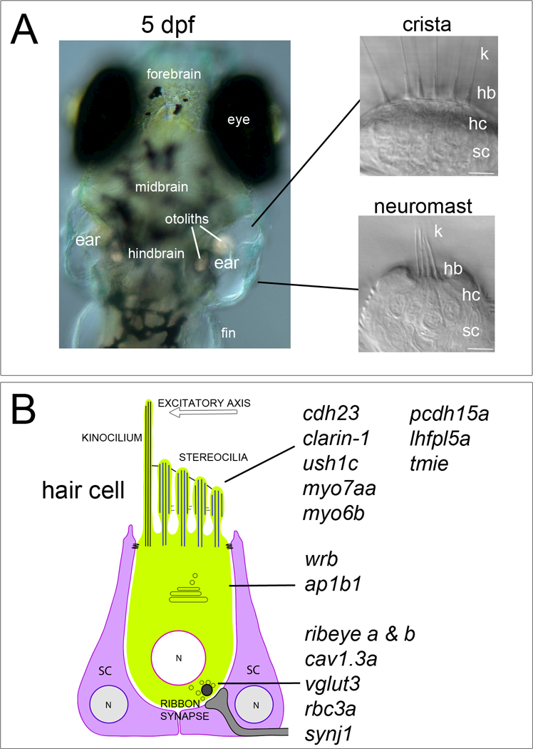
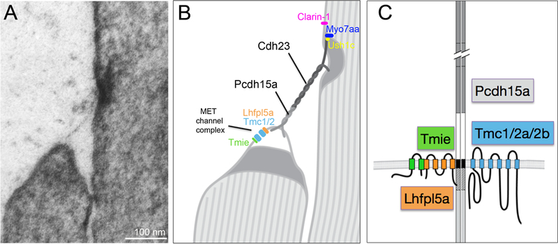
Our ears are remarkable sensory organs, providing the important senses of balance and hearing. The complex structure of the inner ear, or 'labyrinth', along with the assorted neuroepithelia, have evolved to detect head movements and sounds with impressive sensitivity. The rub is that the inner ear is highly vulnerable to genetic lesions and environmental insults. According to National Institute of Health estimates, hearing loss is one of the most commonly inherited or acquired sensorineural diseases. To understand the causes of deafness and balance disorders, it is imperative to understand the underlying biology of the inner ear, especially the inner workings of the sensory receptors. These receptors, which are termed hair cells, are particularly susceptible to genetic mutations - more than two dozen genes are associated with defects in this cell type in humans. Over the past decade, a substantial amount of progress has been made in working out the molecular basis of hair-cell function using vertebrate animal models. Given the transparency of the inner ear and the genetic tools that are available, zebrafish have become an increasingly popular animal model for the study of deafness and vestibular dysfunction. Mutagenesis screens for larval defects in hearing and balance have been fruitful in finding key components, many of which have been implicated in human deafness. This review will focus on the genes that are required for hair-cell function in zebrafish, with a particular emphasis on mechanotransduction. In addition, the generation of new tools available for the characterization of zebrafish hair-cell mutants will be discussed.
Keywords: Hair cell; deafness gene; hair bundle; inner ear; lateral line organ; mechanotransduction; zebrafish.
Figures
References
-
- Ahmed ZM, Goodyear R, Riazuddin S, Lagziel A, Legan PK, Behra M, Burgess SM, Lilley KS, Wilcox ER, Riazuddin S, et al. (2006). The tip-link antigen, a protein associated with the transduction complex of sensory hair cells, is protocadherin-15. J. Neurosci. Off. J. Soc. Neurosci. 26, 7022–7034. - PMC - PubMed
-
- Alsina B, and Whitfield TT (2016). Sculpting the labyrinth: Morphogenesis of the developing inner ear. Semin. Cell Dev. Biol. - PubMed
-
- Avraham KB, Hasson T, Steel KP, Kingsley DM, Russell LB, Mooseker MS, Copeland NG, and Jenkins NA (1995). The mouse Snell’s waltzer deafness gene encodes an unconventional myosin required for structural integrity of inner ear hair cells. Nat. Genet. 11, 369–375. - PubMed
Publication types
MeSH terms
Substances
Grants and funding
LinkOut - more resources
Full Text Sources
Other Literature Sources
Miscellaneous