. 2017 Jul 14;18(1):25.
doi: 10.1186/s12860-017-0142-z.

The M-phase specific hyperphosphorylation of Staufen2 involved the cyclin-dependent kinase CDK1

Affiliations

The M-phase specific hyperphosphorylation of Staufen2 involved the cyclin-dependent kinase CDK1

Rémy Beaujois et al. BMC Cell Biol. .

Erratum in

Abstract

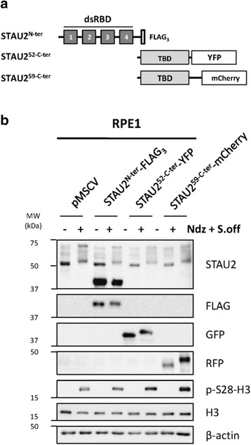
Background: Staufen2 (STAU2) is an RNA-binding protein involved in the post-transcriptional regulation of gene expression. This protein was shown to be required for organ formation and cell differentiation. Although STAU2 functions have been reported in neuronal cells, its role in dividing cells remains deeply uncharacterized. Especially, its regulation during the cell cycle is completely unknown.

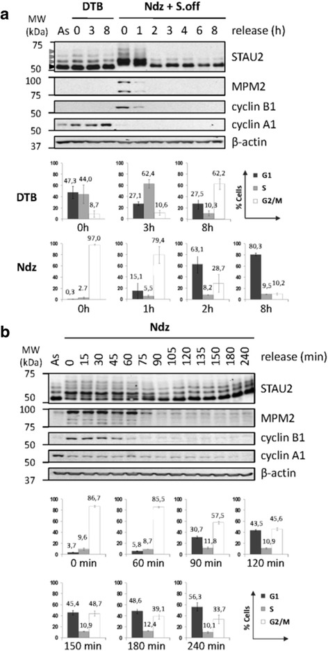
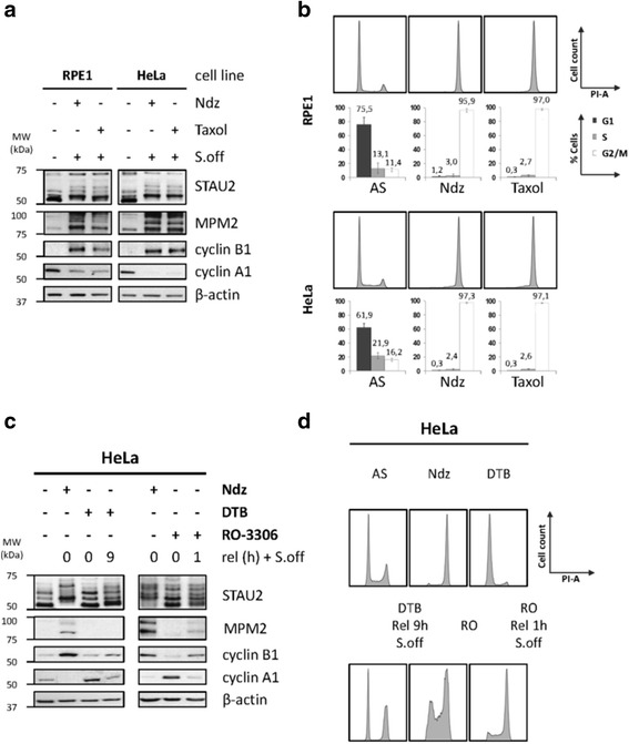
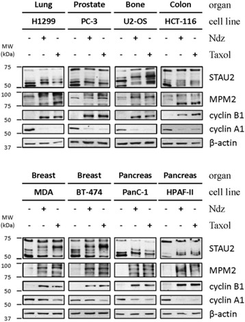
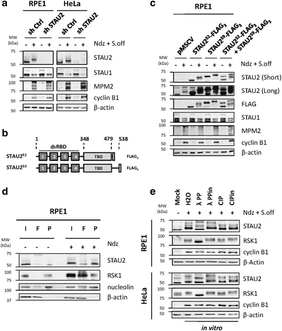
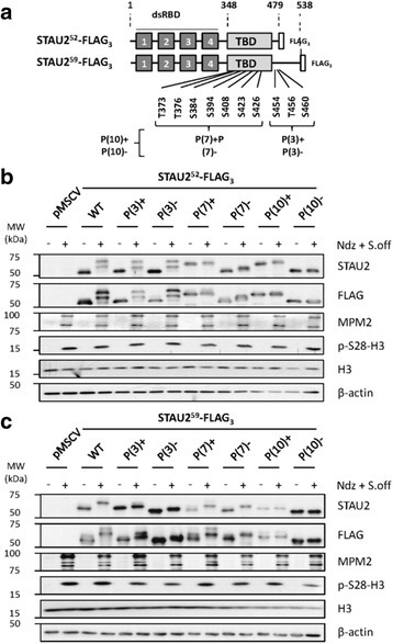
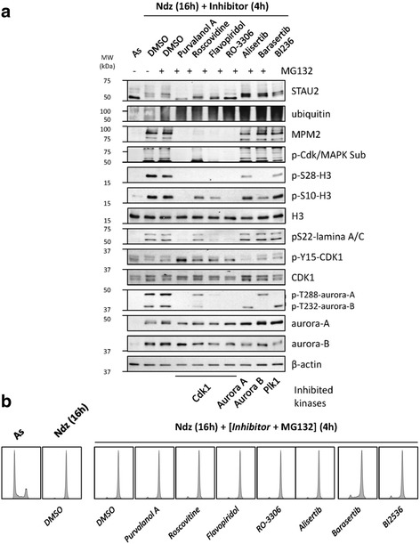
Results: In this study, we showed that STAU2 isoforms display a mitosis-specific slow migration pattern on SDS-gels in all tested transformed and untransformed cell lines. Deeper analyses in hTert-RPE1 and HeLa cells further indicated that the slow migration pattern of STAU2 isoforms is due to phosphorylation. Time course studies showed that STAU2 phosphorylation occurs before prometaphase and terminates as cells exit mitosis. Interestingly, STAU2 isoforms were phosphorylated on several amino acid residues in the C-terminal half via the cyclin-dependent kinase 1 (Cdk1), an enzyme known to play crucial roles during mitosis. Introduction of phospho-mimetic or phospho-null mutations in STAU2 did not impair its RNA-binding capacity, its stability, its interaction with protein co-factors or its sub-cellular localization, suggesting that STAU2 phosphorylation in mitosis does not regulate these functions. Similarly, STAU2 phosphorylation is not likely to be crucial for cell cycle progression since expression of phosphorylation mutants in hTert-RPE1 cells did not impair cell proliferation.

Conclusions: Altogether, these results indicate that STAU2 isoforms are phosphorylated during mitosis and that the phosphorylation process involves Cdk1. The meaning of this post-translational modification is still elusive.

Keywords: Cell cycle; Cyclin-dependent kinase; Mitosis; Phosphorylation; RNA-binding protein; Staufen2.


Conflict of interest statement

Ethics approval and consent to participate

Not applicable.

Consent for publication

Not applicable.

Competing interests

The authors declare that they have no competing interests.

Publisher’s Note

Springer Nature remains neutral with regard to jurisdictional claims in published maps and institutional affiliations.

Figures

Fig. 1
Fig. 1
STAU2 is differentially regulated through the cell cycle in hTert-RPE1 cells. a hTert-RPE1 cells were synchronized by a double thymidine block (DTB) or a nocodazole arrest (Ndz) followed by shake off (S.off). Cells were then released in a fresh medium for different time periods as indicated (Rel (h)). Asynchronous (As) cells were collected as controls. Protein extracts from synchronized cells were analyzed by SDS-PAGE and western blotting to investigate STAU2 pattern migration and expression of cell cycle markers (MPM2 and cyclins). β-actin was used as loading control. As control of synchronization, the percentage of cell population in the G1, S or G2/M phases was determined by FACS analysis (n = 3)(see also Additional file 1). b STAU2 migration dynamics was examined in hTert-RPE1 cells. Cells were blocked in prometaphase with nocodazole (Ndz) without shake-off, released in fresh medium and harvested every 15 min (Rel (min)). Extracts from untreated asynchronous (As) and nocodazole-treated cells were analyzed by western blotting. The percentage of cell population within the G1, S or G2/M phases was determined by FACS (n = 3). In both (a) and (b), Western blots are representatives of three independently performed experiments that showed similar profiles. Error bars represent the standard deviation
Fig. 2
Fig. 2
Post-translational modification of STAU2 in mitosis. a hTert-RPE1 and HeLa cells were synchronized in mitosis with either nocodazole (Ndz) or paclitaxel (Taxol) and enriched by shake off (S.off). Protein extracts from synchronized cells were analyzed by SDS-PAGE and western blotting to investigate STAU2 migration pattern and expression of cell cycle markers (MPM2 and cyclins). β-actin was used as loading control. b As control of synchronization, the percentage of cell population in the G1, S or G2/M phases was determined by FACS analysis (n = 3). c HeLa cells were synchronized either in prometaphase by nocodazole (Ndz), in G1/S transition by double-thymidine block (DTB) or in late G2 by the CDK1 inhibitor RO-3306 (RO-3306) and released from the block for the indicated time periods to reach mitosis. Protein extracts from synchronized cells were analyzed by SDS-PAGE and western blotting to investigate STAU2 migration pattern and expression of mitotic markers (MPM2 and cyclins). β-actin was used as loading control. d As control of synchronization, the percentage of cell population in the G1, S or G2/M phases was determined by FACS analysis (n = 3). Error bars represent the standard deviation
Fig. 3
Fig. 3
Mitosis-specific migration pattern of STAU2 is observed in all tested cell lines. Eight cell lines derived from different organs were synchronized (+) by either nocodazole (Ndz) or paclitaxel (Taxol) and cell extracts were analyzed by western blotting to investigate STAU2 migration pattern and expression of cell cycle markers (MPM2 and cyclins). β-actin was used as loading control. Synchronization was confirmed by FACS (see Additional file 4). Western blots are representatives of three independently performed experiments that showed similar profiles
Fig. 4
Fig. 4
STAU2 is hyperphosphorylated during the M-phase. a shRNA control (shCtrl) or against STAU2 (shSTAU2) were cloned in a retrovirus vector to infect hTert-RPE1 and HeLa cells. Cells were synchronized in prometaphase with nocodazole and mitotic cells were enriched by gentle shake off (Ndz + S.off). Protein extracts from asynchronous (−) and synchronized cells (+) were analyzed by western blotting to detect STAU2 migration and cell cycle markers (MPM2 and cyclins). β-actin was used as loading control. b Schematic representation of STAU2 expression vectors, STAU252-FLAG3 and STAU259-FLAG3. Dark gray boxes, double-stranded RNA-binding domains (dsRBD); light gray boxes, tubulin-binding domain (TBD); white boxes, FLAG3. c hTert-RPE1 cells infected with viruses expressing the empty pMSCV vector (pMSCV), STAU252-FLAG3, STAU259-FLAG3 or both were synchronized (+) by nocodazole and shake off (Ndz + S.off). Migration of STAU2 proteins was detected by SDS-PAGE and western blotting. Both endogenous and overexpressed STAU259-FLAG3 were analyzed with anti-STAU2 antibody, while anti-FLAG antibody was used to specifically recognize FLAG3-tagged STAU2 isoforms. Mitotic marker accumulation was assessed with anti-MPM2 and anti-cyclin B1 antibodies. Loading was normalized with β-actin antibody. d Asynchronous (−) and nocodazole-treated (Ndz)(+) hTert-RPE1 cells were lysed and protein extracts were subjected to separation on phospho-columns. Input from total extracts (I), flow through (F) and phospho-eluates (P) were analyzed by western blotting using anti-STAU2. Anti-RSK1, anti-nucleolin and anti-β-actin antibodies were used as controls for phosphorylated and unphosphorylated proteins, respectively. e Protein extracts from nocodazole-treated (+) hTert-RPE1 and HeLa were incubated in vitro with either water (H2O), Lambda Phosphatase (λPP), inactivated Lambda Phosphatase (λPPin), Calf Intestinal Alkaline Phosphatase (CIP) or inactivated Calf Intestinal Alkaline Phosphatase (CIPin). STAU2 phosphorylation status was analyzed by SDS-PAGE and western blotting. Untreated cells (Mock) were used as control for dephosphorylated STAU2. All western blots are representatives of three independently performed experiments that showed similar profiles
Fig. 5
Fig. 5
STAU2 is phosphorylated in the tubulin-binding domain during mitosis. a Schematic representation of STAU2 deletion mutants. Dark gray boxes, double-stranded RNA-binding domains (dsRBD); light gray boxes, tubulin-binding domain (TBD); white boxes, yellow fluorescent protein (YFP) or red fluorescent protein (mCherry). b hTert-RPE1 cells were infected with retrovirus expressing either STAU2N-ter-FLAG3, STAU252-C-ter-YFP or STAU259-C-ter-mCherry and protein extracts were analyzed by western blotting to detect STAU2 phosphorylation pattern migration (anti-STAU2, anti-FLAG, anti-GFP and anti-RFP) and expression of mitotic markers (p-S28-H3). β-actin was used as loading control. Western blots are representatives of three independently performed experiments that showed similar profiles
Fig. 6
Fig. 6
Multiple residues are phosphorylated in the TBD during mitosis. a Schematic representation of STAU2 showing the 4 double-stranded mRNA-binding domains (dsRBD) and the tubulin-binding domain (TBD). Large-scale mass spectrometry experiments previously identified ten amino acid residues that are specifically phosphorylated during the M-phase. Three, seven and ten residues were simultaneously mutated to generate phospho-mimetic (P(3)+, P(7)+, P(10)+) and phospho-null (P(3)-, P(7)-, P(10)-) STAU252-FLAG3 and STAU259-FLAG3 mutants in the retroviral pMSCV vector. b,c hTert-RPE1 were infected with viruses expressing empty pMSCV, wild type (WT) or phospho-STAU252-FLAG3 (b) or phospho-STAU259-FLAG3 (c) mutants and selected with puromycin. Asynchronous (−) and mitotic (+)(Ndz + S.off) cell extracts were analyzed by western blotting to detect STAU2 migration and mitotic markers (MPM2 and p-S28-H3). All western blots are representatives of three independently performed experiments that showed similar profiles
Fig. 7
Fig. 7
STAU2 is phosphorylated by Cdk1 during mitosis in hTert-RPE1 cells. a hTert-RPE1 cells were incubated with nocodazole (Ndz) for 16 h in the absence (DMSO) or presence of specific kinase inhibitors for 4 h as indicated. Asynchronous (As) cells were used as controls. Cells were treated with the proteasome inhibitor MG132 (+) to keep cells in mitosis. STAU2 migration on SDS-gels was analyzed by western blotting. Cell markers were used to confirm specific inhibition of kinases. b Cell synchronization was confirmed by FACS. Western blots and FACS are representatives of three independently performed experiments that showed similar profiles

Similar articles

Cited by

References

    1. Tyson JJ, Novak B. Temporal organization of the cell cycle. Curr Biol. 2008;18(17):R759–R768. doi: 10.1016/j.cub.2008.07.001. - DOI - PMC - PubMed
    1. Kops GJ, Weaver BA, Cleveland DW. On the road to cancer: aneuploidy and the mitotic checkpoint. Nat Rev Cancer. 2005;5(10):773–785. doi: 10.1038/nrc1714. - DOI - PubMed
    1. Lim S, Kaldis P. Cdks, cyclins and CKIs: roles beyond cell cycle regulation. Dev (Cambridge, England) 2013;140(15):3079–3093. doi: 10.1242/dev.091744. - DOI - PubMed
    1. Malumbres M, Harlow E, Hunt T, Hunter T, Lahti JM, Manning G, et al. Cyclin-dependent kinases: a family portrait. Nat Cell Biol. 2009;11(11):1275–1276. doi: 10.1038/ncb1109-1275. - DOI - PMC - PubMed
    1. Arellano M, Moreno S. Regulation of CDK/cyclin complexes during the cell cycle. Int J Biochem Cell Biol. 1997;29(4):559–573. doi: 10.1016/S1357-2725(96)00178-1. - DOI - PubMed

Publication types

LinkOut - more resources