. 2017 Jul 14;8(1):168.
doi: 10.1186/s13287-017-0618-y.

Interleukin-3 enhances the migration of human mesenchymal stem cells by regulating expression of CXCR4

Affiliations

Interleukin-3 enhances the migration of human mesenchymal stem cells by regulating expression of CXCR4

Amruta Barhanpurkar-Naik et al. Stem Cell Res Ther. .

Abstract

Background: Mesenchymal stem cells (MSCs) represent an important source for cell therapy in regenerative medicine. MSCs have shown promising results for repair of damaged tissues in various degenerative diseases in animal models and also in human clinical trials. However, little is known about the factors that could enhance the migration and tissue-specific engraftment of exogenously infused MSCs for successful regenerative cell therapy. Previously, we have reported that interleukin-3 (IL-3) prevents bone and cartilage damage in animal models of rheumatoid arthritis and osteoarthritis. Also, IL-3 promotes the differentiation of human MSCs into functional osteoblasts and increases their in-vivo bone regenerative potential in immunocompromised mice. However, the role of IL-3 in migration of MSCs is not yet known. In the present study, we investigated the role of IL-3 in migration of human MSCs under both in-vitro and in-vivo conditions.

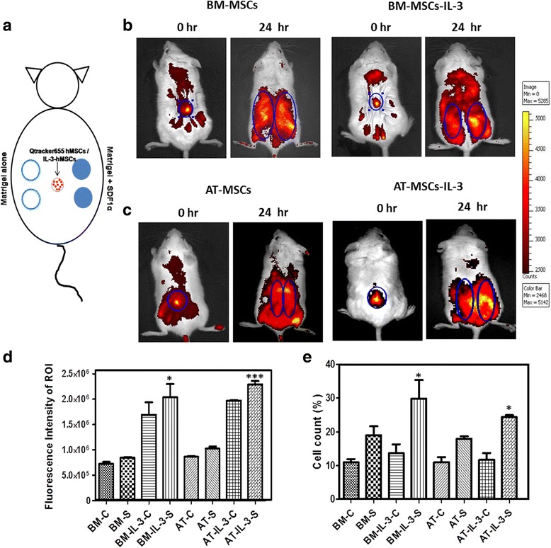
Methods: MSCs isolated from human bone marrow, adipose and gingival tissues were used for in-vitro cell migration, motility and wound healing assays in the presence or absence of IL-3. The effect of IL-3 preconditioning on expression of chemokine receptors and integrins was examined by flow cytometry and real-time PCR. The in-vivo migration of IL-3-preconditioned MSCs was investigated using a subcutaneous matrigel-releasing stromal cell-derived factor-1 alpha (SDF-1α) model in immunocompromised mice.

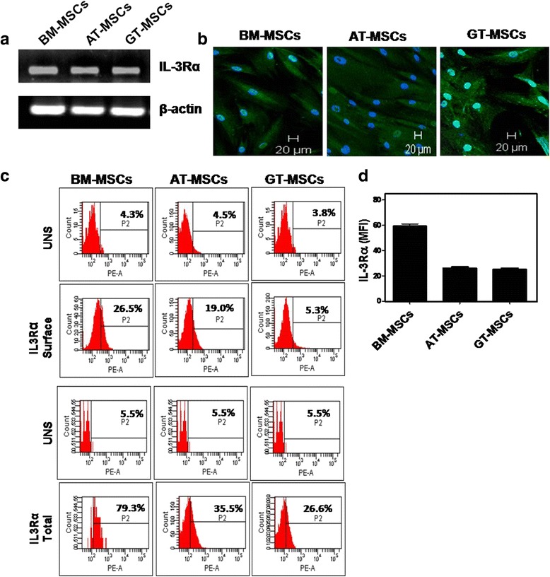
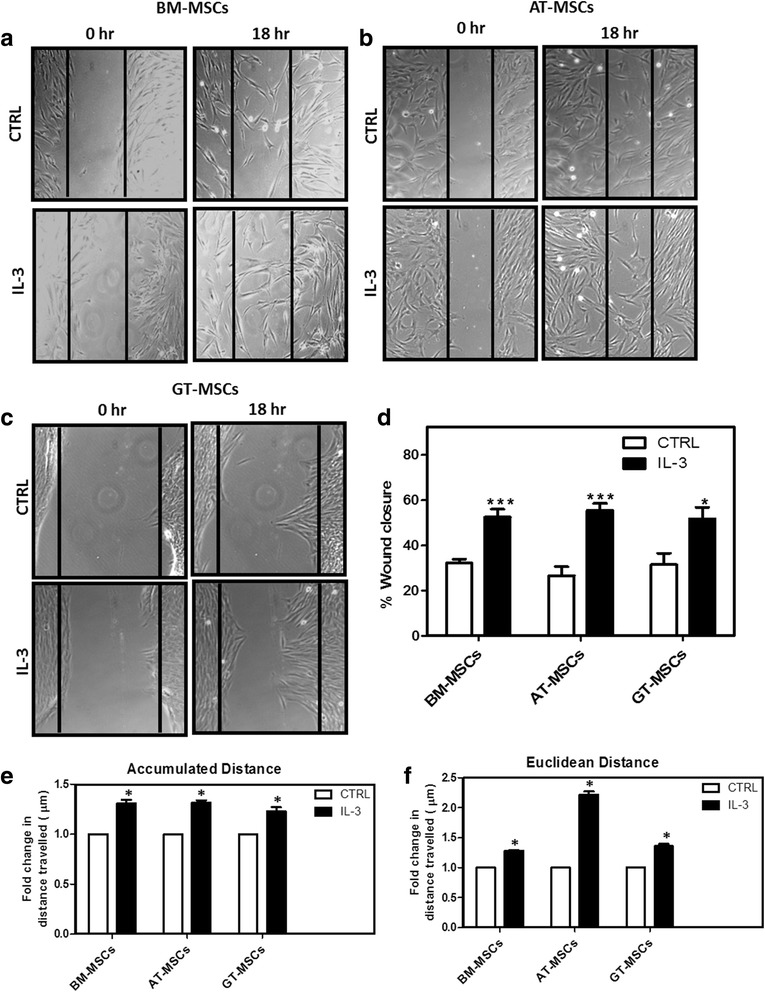
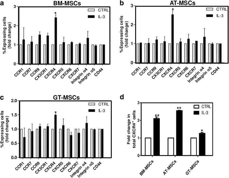
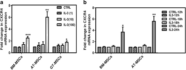
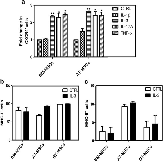
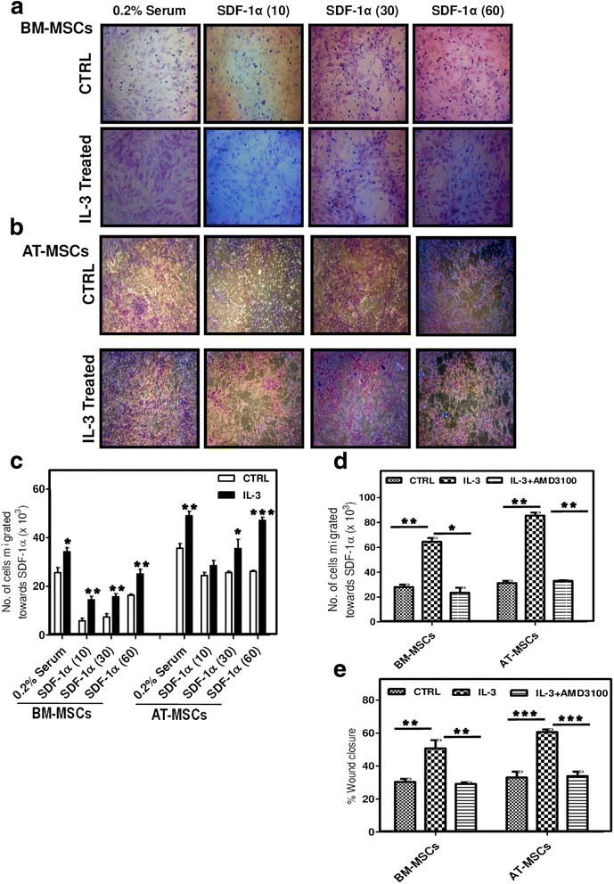
Results: We observed that human MSCs isolated from all three sources express IL-3 receptor-α (IL-3Rα) both at gene and protein levels. IL-3 significantly enhances in-vitro migration, motility and wound healing abilities of MSCs. Moreover, IL-3 preconditioning upregulates expression of chemokine (C-X-C motif) receptor 4 (CXCR4) on MSCs, which leads to increased migration of cells towards SDF-1α. Furthermore, CXCR4 antagonist AMD3100 decreases the migration of IL-3-treated MSCs towards SDF-1α. Importantly, IL-3 also induces in-vivo migration of MSCs towards subcutaneously implanted matrigel-releasing-SDF-1α in immunocompromised mice.

Conclusions: The present study demonstrates for the first time that IL-3 has an important role in enhancing the migration of human MSCs through regulation of the CXCR4/SDF-1α axis. These findings suggest a potential role of IL-3 in improving the efficacy of MSCs in regenerative cell therapy.

Keywords: CXCR4; Cell migration; Interleukin-3; Mesenchymal stem cells; SDF-1α.


Conflict of interest statement

Authors’ information

Not applicable.

Ethics approval and consent to participate

Protocols for animal experiments were approved by the Institutional Animal Ethical Committee (IAEC) of the National Centre for Cell Science, Pune, India (EAF/2014/B-237).

Human bone marrow, adipose tissue and gingival tissue samples were collected from local hospitals after obtaining informed consent with the compliance of the Institutional Ethical Committee (IEC) and the Institutional Committee for Stem Cell Research (IC-SCR) of the National Centre for Cell Science, Pune, India (NCCS/IC-SCR/2013/7).

Consent for publication

Not applicable.

Competing interests

The authors declare that they have no competing interests.

Publisher’s Note

Springer Nature remains neutral with regard to jurisdictional claims in published maps and institutional affiliations.

Figures

Fig. 1
Fig. 1
Human MSCs express IL-3Rα. Human BM-MSCs, AT-MSCs and GT-MSCs of passage 2 were subjected to RT-PCR (a), confocal (b, magnification 10×) and flow cytometry (c) analysis to examine the expression of IL-3Rα both at mRNA and protein levels. Graphical representation of mean fluorescent intensity (MFI) of IL-3Rα on human MSCs (d). Similar results were obtained in two independent experiments. AT adipose tissue, BM bone marrow, GT gingival tissue, IL-3Rα interleukin-3 receptor alpha, MSC mesenchymal stem cell
Fig. 2
Fig. 2
Effect of IL-3 on wound healing and cell motility of human MSCs. BM-MSCs, AT-MSCs and GT-MSCs (104 cells/well) were seeded in 24-well culture plates. After 80–90% confluency, wounds were created on monolayers using a 200 μl pipette tip. Cells were washed and incubated for 18 hours in the absence or presence of human IL-3 (100 ng/ml). Representative images of human BM-MSCs (a), AT-MSCs (b) and GT-MSCs (c) at 0 and 18 hours of IL-3 treatment (Magnification 10×). Percent wound closure from three independent experiments was analyzed (d). Human BM-MSCs, AT-MSCs and GT-MSCs were incubated for 24 hours with and without IL-3 and cell motility was examined by measurement of accumulated (e) and euclidean (f) distance travelled by MSCs. The cell motility images were captured by time-lapse microscope and analyzed using Image J Software. Data shown as mean ± SEM of three independent experiments. *p ≤ 0.05 and ***p ≤ 0.001 vs control group. AT adipose tissue, BM bone marrow, CTRL control, GT gingival tissue, IL-3 interleukin-3, MSC mesenchymal stem cell
Fig. 3
Fig. 3
Effect of IL-3 on expression of chemokine receptors and integrins involved in migration of MSCs. BM-MSCs, AT-MSCs and GT-MSCs were treated with IL-3 (100 ng/ml) for 24 hours and surface expression of different chemokine receptors and integrins was analyzed by flow cytometry. ac Fold change as percentage of cells expressing chemokine receptors and integrins. d Fold change in percentage of cells expressing intracellular CXCR4 analyzed after IL-3 treatment in permeabilized MSCs. Data shown as mean ± SEM of three independent experiments. *p ≤ 0.05 and **p ≤ 0.01 vs control groups. AT adipose tissue, BM bone marrow, CTRL control, GT gingival tissue, IL-3 interleukin-3, MSC mesenchymal stem cell
Fig. 4
Fig. 4
Effect of IL-3 on mRNA expression of CXCR4 in MSCs. BM-MSCs, AT-MSCs and GT-MSCs were treated with different concentrations of IL-3 for 24 hours and fold change in mRNA expression of CXCR4 was analyzed using real-time PCR (a). CXCR4 mRNA expression in MSCs incubated with IL-3 (100 ng/ml) was analyzed after 12, 18 and 24 hours (b). Data shown as mean ± SEM of three independent experiments. *p ≤ 0.05, **p ≤ 0.01 and ***p ≤ 0.001 vs control groups. AT adipose tissue, BM bone marrow, CTRL control, CXCR4 chemokine (C-X-C motif) receptor 4, GT gingival tissue, IL-3 interleukin-3, MSC mesenchymal stem cell
Fig. 5
Fig. 5
Comparison of effect of different cytokines on regulation of CXCR4. To compare the induction of CXCR4 expression by different cytokines, BM-MSCs and AT-MSCs were incubated with IL-1β (10 ng/ml), IL-3 (100 ng/ml), IL-17A (50 ng/ml) and TNF-α (50 ng/ml) independently for 24 hours. Fold change in percentage of CXCR4+ cells was analyzed by flow cytometry (a). Expression of MHC class I (b) and class II (c) molecules on MSCs was analyzed by flow cytometry after IL-3 treatment for 24 hours, and compared with respective untreated MSCs. Data shown as mean ± SEM of three independent experiments. *p ≤ 0.05 and **p ≤ 0.01 vs control groups. AT adipose tissue, BM bone marrow, CTRL control, CXCR4 chemokine (C-X-C motif) receptor 4, GT gingival tissue, IL-3 interleukin-3, MSC mesenchymal stem cell
Fig. 6
Fig. 6
IL-3-treated MSCs migrate towards SDF-1α. IL-3-pretreated (100 ng/ml) and untreated BM-MSCs and AT-MSCs were seeded in the upper chamber of cell culture inserts and SDF-1α (10, 30, 60 ng/ml) was added to the lower chamber. After 18 hours, migration of cells towards SDF-1α was visualized by staining the cells from the lower side of inserts with hematoxylin (a, b). Cells from the lower side were also removed by trypsinization and counted (c). IL-3-pretreated and untreated BM-MSCs and AT-MSCs were seeded in the upper chamber of cell culture inserts with or without AMD3100 (10 μM) and SDF-1α (60 ng/ml) was added to the lower chamber. After 18 hours, cells from the lower side of inserts were trypsinized and counted (d). Wounds created on monolayers of BM-MSCs and AT-MSCs were incubated with IL-3 in the presence or absence of AMD3100 and percent wound closure was analyzed (e). Data are representative of two independent experiments. *p ≤ 0.05, **p ≤ 0.01 and***p ≤ 0.001 vs control group or vs IL-3-pretreated group. AT adipose tissue, BM bone marrow, CTRL control, IL-3 interleukin-3, MSC mesenchymal stem cell, SDF-1α stromal cell-derived factor-1 alpha
Fig. 7
Fig. 7
In-vivo migration of IL-3-treated cells towards SDF-1α. Two matrigel plugs mixed with SDF-1α (100 ng/ml) were injected subcutaneously at the right dorsal side of NOD/SCID mice and two matrigel plugs without SDF-1α were injected on the left dorsal side of mice. BM-MSCs and AT-MSCs untreated or pretreated with IL-3 were labeled with Qtracker 655 and injected subcutaneously (105 cells in 100 μl) at the center, equidistant from all four implants. Schematic representation of the subcutaneously implanted matrigel in mouse model (a). Mice were acquired on the Live Cell Imaging System at 0 hours for detection of labeled MSCs and at 24 hours for detection of migrated MSCs towards the matrigel plugs (b, c). Graphical representation showing the counts of fluorescent intensity at the region of interest (ROI), the area of matrigel plugs (d). Difference between in-vivo migration of IL-3-treated and untreated MSCs towards SDF-1α (BM-IL-3-S vs BM-S and AT-IL-3-S vs AT-S) was compared. Matrigel plugs were harvested from mice and isolated cells were acquired on flow cytometry (e). C matrigel implant without SDF-1α, S matrigel implant containing SDF-1α. Data shown as mean ± SEM (mice, n = 6 and matrigel plugs, n = 12/group). *p ≤ 0.05 and ***p ≤ 0.001 vs control groups. AT adipose tissue, BM bone marrow, IL-3 interleukin-3, MSC mesenchymal stem cell, SDF-1α stromal cell-derived factor-1 alpha

References

    1. Uccelli A, Moretta L, Pistoia V. Mesenchymal stem cells in health and disease. Nat Rev Immunol. 2008;8:726–36. doi: 10.1038/nri2395. - DOI - PubMed
    1. Djouad F, Bouffi C, Ghannam S, Noel D, Jorgensen C. Mesenchymal stem cells: innovative therapeutic tools for rheumatic diseases. Nat Rev Rheumatol. 2009;5:392–9. doi: 10.1038/nrrheum.2009.104. - DOI - PubMed
    1. Chamberlain G, Fox J, Ashton B, Middlenton J. Concise review: mesenchymal stem cells: their phenotype, differentiation capacity, immunological features, and potential for homing. Stem Cells. 2007;25:2739–49. doi: 10.1634/stemcells.2007-0197. - DOI - PubMed
    1. Ma S, Xie N, Li W, Yuan B, Shi Y, Wang Y. Immunobiology of mesenchymal stem cells. Cell Death Differ. 2014;21:216–25. doi: 10.1038/cdd.2013.158. - DOI - PMC - PubMed
    1. Barbash IM, Chouraqui P, Baron J, Feinberg MS, Etzion S, Tessone A, et al. Systemic delivery of bone marrow-derived mesenchymal stem cells to the infarcted myocardium: feasibility, cell migration, and body distribution. Circulation. 2003;108:863–68. doi: 10.1161/01.CIR.0000084828.50310.6A. - DOI - PubMed