. 2017 Oct 1;313(4):G330-G341.
doi: 10.1152/ajpgi.00161.2017. Epub 2017 Jul 13.

Augmentation of cGMP/PKG pathway and colonic motility by hydrogen sulfide

Affiliations

Augmentation of cGMP/PKG pathway and colonic motility by hydrogen sulfide

Ancy D Nalli et al. Am J Physiol Gastrointest Liver Physiol. .

Abstract

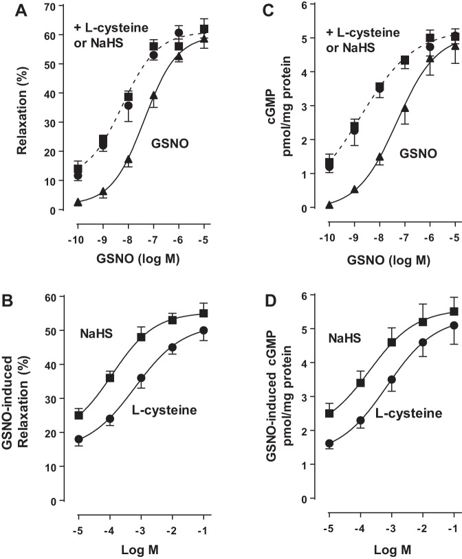
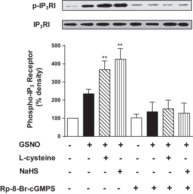
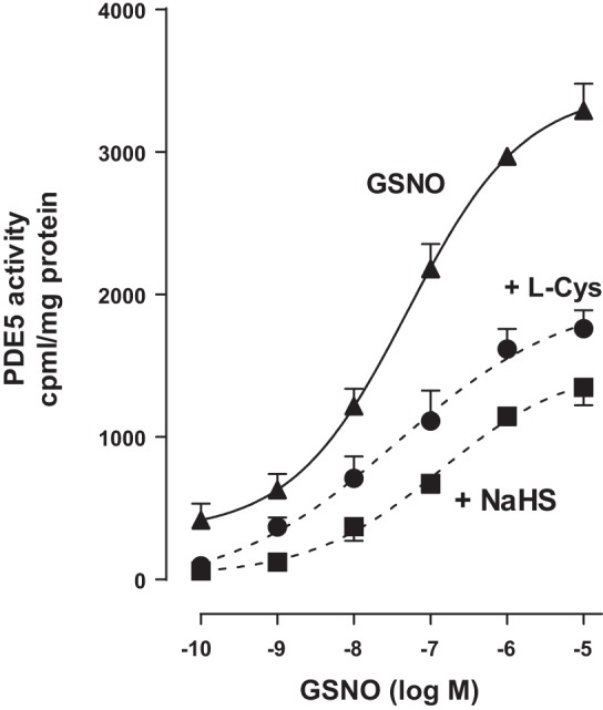
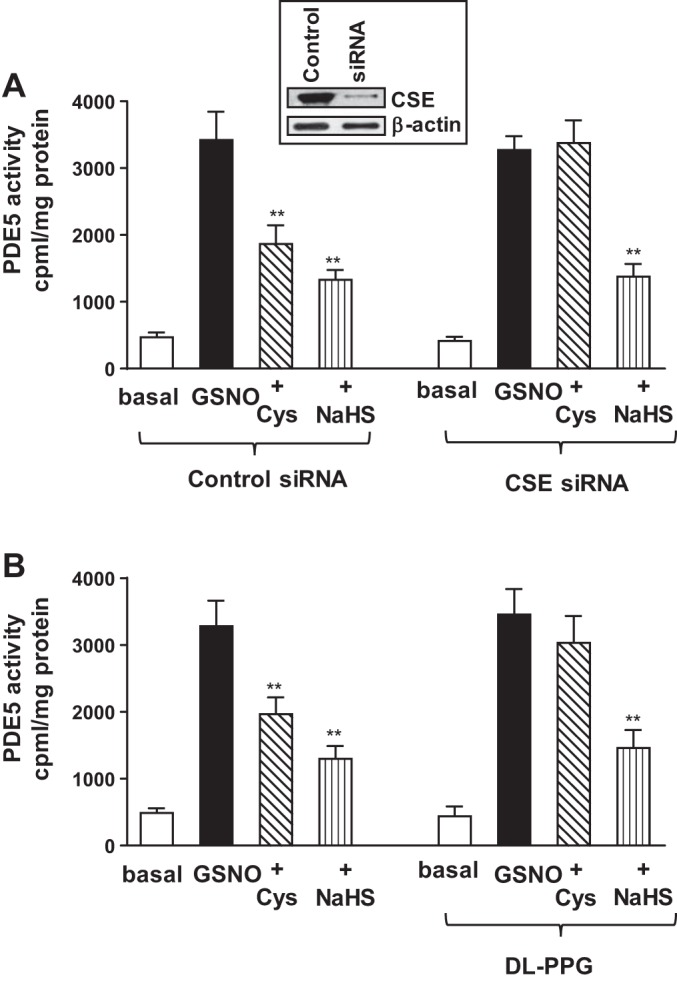
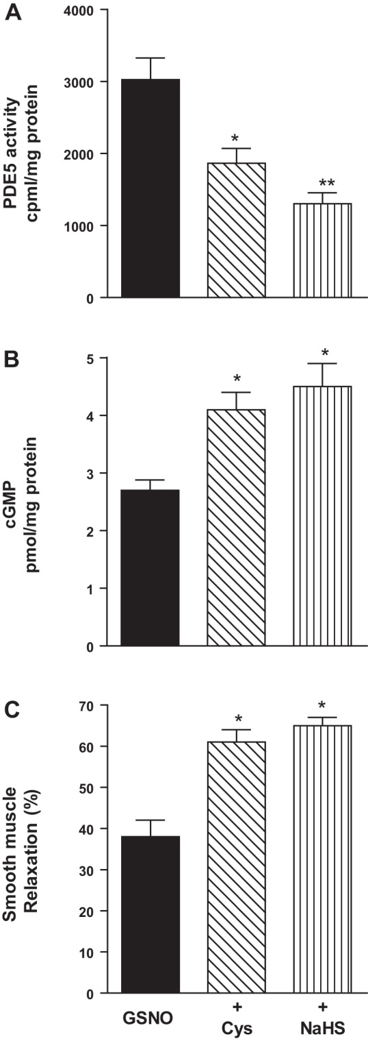
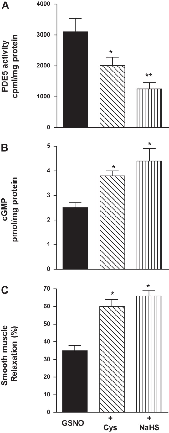
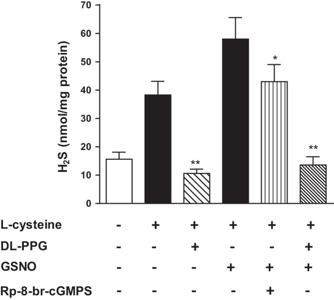
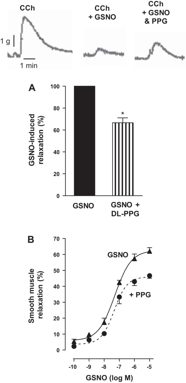
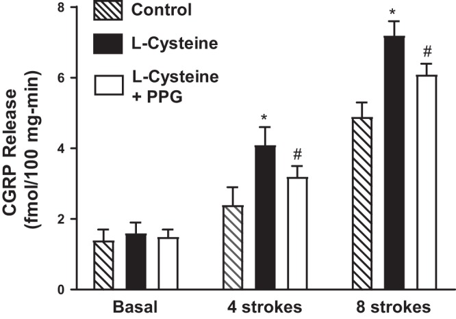
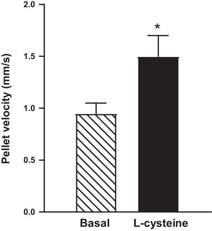
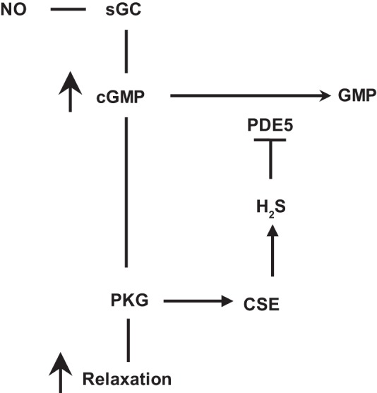
Hydrogen sulfide (H2S), like nitric oxide (NO), causes smooth muscle relaxation, but unlike NO, does not stimulate soluble guanylyl cyclase (sGC) activity and generate cyclic guanosine 5'-monophosphate (cGMP). The aim of this study was to investigate the interplay between NO and H2S in colonic smooth muscle. In colonic smooth muscle from rabbit, mouse, and human, l-cysteine, substrate of cystathionine-γ-lyase (CSE), or NaHS, an H2S donor, inhibited phosphodiesterase 5 (PDE5) activity and augmented the increase in cGMP levels, IP3 receptor phosphorylation at Ser1756 (measured as a proxy for PKG activation), and muscle relaxation in response to NO donor S-nitrosoglutathione (GSNO), suggesting augmentation of cGMP/PKG pathway by H2S. The inhibitory effect of l-cysteine, but not NaHS, on PDE5 activity was blocked in cells transfected with CSE siRNA or treated with CSE inhibitor d,l-propargylglycine (dl-PPG), suggesting activation of CSE and generation of H2S in response to l-cysteine. H2S levels were increased in response to l-cysteine, and the effect of l-cysteine was augmented by GSNO in a cGMP-dependent protein kinase-sensitive manner, suggesting augmentation of CSE/H2S by cGMP/PKG pathway. As a result, GSNO-induced relaxation was inhibited by dl-PPG. In flat-sheet preparation of colon, l-cysteine augmented calcitonin gene-related peptide release in response to mucosal stimulation, and in intact segments, l-cysteine increased the velocity of pellet propulsion. These results demonstrate that in colonic smooth muscle, there is a novel interplay between NO and H2S. NO generates H2S via cGMP/PKG pathway, and H2S, in turn, inhibits PDE5 activity and augments NO-induced cGMP levels. In the intact colon, H2S promotes colonic transit.NEW & NOTEWORTHY Hydrogen sulfide (H2S) and nitric oxide (NO) are important regulators of gastrointestinal motility. The studies herein provide the cross talk between NO and H2S signaling to mediate smooth muscle relaxation and colonic transit. H2S inhibits phosphodiesterase 5 activity to augment cGMP levels in response to NO, which, in turn, via cGMP/PKG pathway, generates H2S. These studies suggest that interventions targeted at restoring NO and H2S homeostasis within the smooth muscle may provide novel therapeutic approaches to mitigate motility disorders.

Keywords: muscle relaxation; nitric oxide; phosphodiesterase 5; signaling.


Figures

Fig. 1.
Fig. 1.
Augmentation of S-nitrosoglutathione (GSNO)-induced muscle relaxation and cyclic guanosine 5′-monophosphate (cGMP) formation by l-cysteine and NaHS in colonic smooth muscle. A: dispersed muscle cells from rabbit colon were treated with increasing concentrations of GSNO (0.1 nM to 10 µM) for 10 min and then treated with carbachol (CCh, 1 µM) for 30 s. In some experiments, cells were cotreated with GSNO and l-cysteine (10 mM) or NaHS (1 mM) for 10 min and then treated with CCh (0.1 µM) for 30 s. B: dispersed muscle cells from rabbit colon were treated with submaximal concentrations of GSNO (10 nM) in the presence or absence of different concentrations of l-cysteine (0.1 µM to 100 mM) or NaHS (0.1 µM to 100 mM) for 10 min and then treated with CCh (1 µM) for 30 s. Muscle cell length was measured by scanning micrometry. Contraction by CCh was calculated as a percent decrease in muscle cell length from control cell length (35 ± 5% decrease in cell length from the basal cell length 97 ± 5 µm). Relaxation in response to GSNO in the presence or absence of l-cysteine or NaHS was expressed as the percent decrease in CCh-induced contraction. Values are means ± SE of four experiments. C: dispersed muscle cells from rabbit colon were treated with increasing concentrations of GSNO (0.1 nM to 10 µM) in the presence or absence of l-cysteine (10 mM) or NaHS (1 mM) for 10 min. D: dispersed muscle cells from rabbit colon were treated with submaximal concentrations of GSNO (10 nM) in the presence or absence of different concentration of l-cysteine (0.1 to 100 mM) or NaHS (0.1 µM to 100 mM) for 10 min. cGMP levels were measured by radioimmunoassay using [125I]cGMP. Results are expressed as picomole per milligrams of protein. Values are means ± SE of five experiments.
Fig. 2.
Fig. 2.
Augmentation of GSNO-induced PKG activity (IP3 receptor phosphorylation) by l-cysteine and NaHS in colonic smooth muscle. Dispersed muscle cells from rabbit colon were treated with GSNO (10 nM) in the presence or absence of l-cysteine (10 mM) or NaHS (1 mM) for 10 min. In some experiments, cells were pretreated with PKG inhibitor Rp-8-Br-cGMPS (10 µM) before the addition of GSNO in the presence or absence of l-cysteine or NaHS. IP3 receptor phosphorylation at Ser1756, a proxy for PKG activity, was measured using phospho-specific (Ser1756) antibody. Results are expressed as percent density of phosphorylated protein bands. Values are means ± SE of four experiments. Representative images of four separate experiments were shown in the figure. **P < 0.01, significant augmentation of GSNO-induced IP3 receptor phosphorylation.
Fig. 3.
Fig. 3.
Inhibition of GSNO-stimulated PDE5 activity by l-cysteine and NaHS. Dispersed rabbit colonic smooth muscle cells were treated with GSNO (0.1 nM to 10 µM) for 10 min. PDE5 was immunoprecipitated from lysates of muscle cells, and PDE5 activity was measured using [3H]cGMP as the substrate. In some experiments, muscle cells were pretreated with l-cysteine (10 mM) and NaHS (1 mM) for 10 min followed by treatment with GSNO. The increase in PDE5 activity in response to GSNO was measured by an increase in [3H]GMP levels in the effluent, and the results are expressed as counts/min per milligram protein above basal [3H]GMP levels (264 ± 43 cpm/mg protein). Values are means ± SE of four experiments.
Fig. 4.
Fig. 4.
Inhibition of GSNO-stimulated PDE5 activity by l-cysteine via activation of cystathionine-γ-lyase (CSE). A: cultured rabbit colonic muscle cells were transfected with control siRNA or CSE-specific siRNA for 48 h. Cells were treated with GSNO (10 µM) for 10 min in the presence or absence of l-cysteine (10 mM) or NaHS (1 mM) pretreatment for 10 min. PDE5 was immunoprecipitated from lysates of muscle cells, and PDE5 activity was measured using [3H]cGMP as the substrate. The increase in PDE5 activity in response to GSNO was measured by an increase in [3H]GMP levels in the effluent, and the results are expressed as counts/min per milligram protein. Values are means ± SE of four experiments. **P < 0.01, significant inhibition of GSNO-induced PDE5 activity. Expression of CSE in cells transfected with control siRNA or CSE siRNA was analyzed by Western blot analysis (inset). B: dispersed muscle cells from rabbit colon were treated with GSNO (10 µM) for 10 min in the presence or absence of l-cysteine (10 mM) or NaHS (1 mM) pretreatment of 10 min. In some experiments, cells were treated with l-cysteine (10 mM) or NaHS (1 mM) in the presence of CSE inhibitor dl-PPG (1 mM). PDE5 was immunoprecipitated from lysates of muscle cells, and PDE5 activity was measured using [3H]cGMP as the substrate. The increase in PDE5 activity in response to GSNO was measured by an increase in [3H]GMP levels in the effluent, and the results are expressed as counts/min per milligram protein. Values are means ± SE of four experiments. **P < 0.01, significant inhibition of GSNO-induced PDE5 activity.
Fig. 5.
Fig. 5.
Inhibition of GSNO-induced PDE5 activity and augmentation of cGMP formation and relaxation by l-cysteine and NaHS in mouse colonic muscle cells. A: dispersed mouse colonic smooth muscle cells were treated with GSNO (10 µM) for 10 min in the presence or absence of with l-cysteine (10 mM) and NaHS (1 mM) pretreatment for 10 min. PDE5 was immunoprecipitated from lysates of muscle cells, and PDE5 activity was measured using [3H]cGMP as the substrate. The increase in PDE5 activity in response to GSNO was measured by an increase in [3H]GMP levels in the effluent, and the results are expressed as counts/min per milligram protein above basal [3H]GMP levels (238 ± 36 cpm/mg protein). Values are means ± SE of four experiments. *P < 0.05, **P < 0.01 significant inhibition of GSNO-induced PDE5 activity. B: dispersed muscle cells from mouse colon were treated with submaximal concentrations of GSNO (100 nM) for 10 min in the presence or absence of l-cysteine (10 mM) or NaHS (1 mM) pretreatment for 10 min. cGMP levels were measured by radioimmunoassay using [125I]cGMP. Results are expressed as picomole per milligrams of protein. Values are means ± SE of five experiments. *P < 0.05, significant augmentation of GSNO-induced cGMP levels. C: dispersed muscle cells from mouse colon were treated with GSNO (100 nM) in the presence or absence of l-cysteine (10 mM) or NaHS (1 mM) for 10 min and then treated with CCh (1 µM) for 30 s. Muscle cell length was measured by scanning micrometry. Contraction by CCh was calculated as a percent decrease in muscle length from control cell length (32 ± 4% decrease in cell length from the basal cell length 112 ± 7 µm). Relaxation in response to GSNO in the presence or absence of l-cysteine or NaHS was expressed as the percent decrease in CCh-induced contraction. Values are means ± SE of four or five experiments. *P < 0.05, significant augmentation of GSNO-induced relaxation.
Fig. 6.
Fig. 6.
Inhibition of GSNO-induced PDE5 activity and augmentation of cGMP formation and relaxation by l-cysteine and NaHS in human colonic muscle cells. A: dispersed human colonic smooth muscle cells were treated with GSNO (10 µM) for 10 min in the presence or absence of with l-cysteine (10 mM) and NaHS (1 mM) pretreatment for 10 min. PDE5 was immunoprecipitated from lysates of muscle cells, and PDE5 activity was measured using [3H]cGMP as the substrate. The increase in PDE5 activity in response to GSNO was measured by an increase in [3H]GMP levels in the effluent, and the results are expressed as counts/min per milligram protein above basal [3H]GMP levels (286 ± 45 cpm/mg protein). Values are means ± SE of four experiments. *P < 0.05, **P < 0.01 significant inhibition of GSNO-induced PDE5 activity. B: dispersed muscle cells from human colon were treated with GSNO (100 nM) for 10 min in the presence or absence of l-cysteine (10 mM) or NaHS (1 mM) pretreatment for 10 min. cGMP levels were measured by radioimmunoassay using [125I]cGMP. Results are expressed as picomole per milligrams of protein. Values are means ± SE of five experiments. *P < 0.05, significant augmentation of GSNO-induced cGMP formation. C: dispersed muscle cells from human colon were treated with GSNO (100 nM) in the presence or absence of l-cysteine (10 mM) or NaHS (1 mM) for 10 min and then treated with CCh (1 µM) for 30 s. Muscle cell length was measured by scanning micrometry. Contraction by CCh was calculated as a percent decrease in muscle length from control cell length (32 ± 4% decrease in cell length from the basal cell length 95 ± 4 µm). Relaxation in response to GSNO in the presence or absence of l-cysteine or NaHS was expressed as the percent decrease in CCh-induced contraction. Values are means ± SE of four experiments. *P < 0.05, significant augmentation of in GSNO-induced relaxation.
Fig. 7.
Fig. 7.
Stimulation of CSE activity in response to l-cysteine and GSNO in colonic smooth muscle. Homogenates prepared from rabbit colonic smooth muscle were treated with l-cysteine (10 mM) for 90 min in the presence or absence of CSE inhibitor d,l-propargylglycine (dl-PPG, 1 mM). In some experiments, homogenates were treated with l-cysteine (10 mM) and GSNO (10 µM) in the presence or absence of PKG inhibitor Rp-8-Br-cGMPS (10 µM). Formation of H2S was measured by spectrophotometry and expressed as nanomole per milligrams tissue. Values are means ± SE of seven experiments. **P < 0.01, significant inhibition of l-cysteine- or GSNO-induced H2S production by dl-PPG; *P < 0.05, significant inhibition of GSNO-induced H2S production by Rp-8-Br-cGMPS.
Fig. 8.
Fig. 8.
Partial reversal of NO-induced relaxation by CSE inhibitor d,l-propargylglycine (dl-PPG) in colonic smooth muscle. A: muscle strips from the rabbit colon were allowed to equilibrate at a passive tension of 1 g for 30 min and then treated with CCh (10 µM). In some experiments, the strips are incubated with GSNO (10 µM) in the presence or absence of CSE inhibitor dl-PPG (1 mM) for 10 min. Relaxation was measured as a percent decrease of contraction (measured as maximal area under the curve induced by CCh) in response to GSNO in the presence or absence of dl-PPG. Values are expressed as percent maximum of GSNO-induced relaxation. Values are means ± SE of four experiments. *P < 0.05, significant inhibition of GSNO-induced relaxation by dl-PPG. Contractile recording shows the inhibition of CCh-induced contraction by GSNO and partial reversal of GSNO-induced inhibition by dl-PPG. B: dispersed muscle cells from rabbit colon were treated with different concentrations of GSNO (0.1 nM to 10 µM) in the presence or absence of dl-PPG (1 mM) for 10 min and then treated with CCh (1 µM) for 30 s. Muscle cell length was measured by scanning micrometry. Contraction by CCh was calculated as a percent decrease in muscle length from control cell length (35 ± 5% decrease in cell length from the basal cell length 102 ± 4 µm). Relaxation in response to GSNO in the presence or absence of dl-PPG was expressed as the percent decrease in CCh-induced contraction. Values are means ± SE of four experiments.
Fig. 9.
Fig. 9.
Increase in calcitonin gene-related peptide (CGRP) release. CGRP release in the central compartment following mucosal stimulation in the presence or absence of l-cysteine (10 mM) alone or l-cysteine plus dl-PPG (10 mM) was measured as described in materials and methods. Basal release represents control release in the absence of mucosal stimulation. Release in the presence of l-cysteine was represented by solid bars. Values are means ± SE of three experiments. *P < 0.05, significant increase in release in the presence of l-cysteine. #P < 0.05, significant inhibition of l-cysteine effect by dl-PPG.
Fig. 10.
Fig. 10.
Velocity of propulsion of fecal pellets in colonic segments from mouse colon. Artificial fecal pellets were inserted into the orad end of intact segments isolated from mouse colon, and the time to traverse a defined distance recorded. The velocity of propulsion of five pellets was calculated. Consistent with augmentation of relaxation of smooth muscle, the velocity of fecal pellet propulsion was increased by 10 mM l-cysteine. Values are mean ± SE of four experiments. *P < 0.05 significant augmentation of colonic propulsion by l-cysteine.
Fig. 11.
Fig. 11.
The interplay between NO and H2S in colonic smooth muscle. In colonic smooth muscle, relaxation in response to nitric oxide (NO) is mediated via stimulation of soluble guanylyl cyclase (sGC) activity, cGMP generation, and cGMP-dependent protein kinase (PKG) activity. PKG phosphorylates various targets in the contractile pathway to decrease intracellular Ca2+ and/or increase myosin light chain phosphatase activity, key requirements in muscle relaxation. cGMP/PKG pathway, in addition, activates cystathionine-γ-lyase (CSE) to generate H2S, which in turn inhibits PDE5 activity and augments cGMP/PKG pathway.

Similar articles

Cited by

References

    1. Abe K, Kimura H. The possible role of hydrogen sulfide as an endogenous neuromodulator. J Neurosci 16: 1066–1071, 1996. - PMC - PubMed
    1. Ali MY, Ping CY, Mok YY, Ling L, Whiteman M, Bhatia M, Moore PK. Regulation of vascular nitric oxide in vitro and in vivo; a new role for endogenous hydrogen sulphide? Br J Pharmacol 149: 625–634, 2006. doi:10.1038/sj.bjp.0706906. - DOI - PMC - PubMed
    1. Beavo JA. Cyclic nucleotide phosphodiesterases: functional implications of multiple isoforms. Physiol Rev 75: 725–748, 1995. - PubMed
    1. Bucci M, Papapetropoulos A, Vellecco V, Zhou Z, Zaid A, Giannogonas P, Cantalupo A, Dhayade S, Karalis KP, Wang R, Feil R, Cirino G. cGMP-dependent protein kinase contributes to hydrogen sulfide-stimulated vasorelaxation. PLoS One 7: e53319, 2012. doi:10.1371/journal.pone.0053319. - DOI - PMC - PubMed
    1. Bucci M, Papapetropoulos A, Vellecco V, Zhou Z, Pyriochou A, Roussos C, Roviezzo F, Brancaleone V, Cirino G. Hydrogen sulfide is an endogenous inhibitor of phosphodiesterase activity. Arterioscler Thromb Vasc Biol 30: 1998–2004, 2010. doi:10.1161/ATVBAHA.110.209783. - DOI - PubMed

LinkOut - more resources