Pacemakers in MRI for the Neuroradiologist
- PMID: 28705821
- PMCID: PMC7963729
- DOI: 10.3174/ajnr.A5314
Pacemakers in MRI for the Neuroradiologist
Abstract
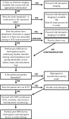
Cardiac implantable electronic devices are frequently encountered in clinical practice in patients being screened for MR imaging examinations. Traditionally, the presence of these devices has been considered a contraindication to undergoing MR imaging. Growing evidence suggests that most of these patients can safely undergo an MR imaging examination if certain conditions are met. This document will review the relevant cardiac implantable electronic devices encountered in practice today, the background physics/technical factors related to scanning these devices, the multidisciplinary screening protocol used at our institution for scanning patients with implantable cardiac devices, and our experience in safely performing these examinations since 2010.
© 2017 by American Journal of Neuroradiology.
Figures


Comment in
-
Reply.AJNR Am J Neuroradiol. 2018 Feb;39(2):E37. doi: 10.3174/ajnr.A5467. Epub 2017 Oct 26. AJNR Am J Neuroradiol. 2018. PMID: 29074630 Free PMC article. No abstract available.
-
Economic Considerations in MR Imaging of Patients with Cardiac Devices.AJNR Am J Neuroradiol. 2018 Feb;39(2):E36. doi: 10.3174/ajnr.A5450. Epub 2017 Oct 26. AJNR Am J Neuroradiol. 2018. PMID: 29074631 Free PMC article. No abstract available.
-
Reply.AJNR Am J Neuroradiol. 2018 May;39(5):E56. doi: 10.3174/ajnr.A5575. Epub 2018 Mar 1. AJNR Am J Neuroradiol. 2018. PMID: 29496727 Free PMC article. No abstract available.
-
Pacemakers in MRI for the Neuroradiologist: Revisited.AJNR Am J Neuroradiol. 2018 May;39(5):E54-E55. doi: 10.3174/ajnr.A5565. Epub 2018 Mar 1. AJNR Am J Neuroradiol. 2018. PMID: 29496728 Free PMC article. No abstract available.
References
-
- OECD. Magnetic resonance imaging (MRI) exams, total. Health: Key Tables. OECD 2014. https://data.oecd.org/healthcare/magnetic-resonance-imaging-mri-exams.htm. Accessed December 24, 2016.
-
- Levine GN, Gomes AS, Arai AE, et al. ; American Heart Association Committee on Diagnostic and Interventional Cardiac Catheterization, American Heart Association Council on Clinical Cardiology, American Heart Association Council on Cardiovascular Radiology and Intervention. Safety of magnetic resonance imaging in patients with cardiovascular devices: an American Heart Association scientific statement from the Committee on Diagnostic and Interventional Cardiac Catheterization, Council on Clinical Cardiology, and the Council on Cardiovascular Radiology and Intervention: endorsed by the American College of Cardiology Foundation, the North American Society for Cardiac Imaging, and the Society for Cardiovascular Magnetic Resonance. Circulation 2007;116:2878–91 10.1161/CIRCULATIONAHA.107.187256 - DOI - PubMed
Publication types
MeSH terms
Grants and funding
LinkOut - more resources
Full Text Sources
Other Literature Sources
Medical
Miscellaneous
